Figure 2. Multimodal imaging evaluation and visual fields, at baseline. A: Color fundus photography reveals small white patches of atrophy around the fovea in the right eye and a well-circumscribed
ring-shaped area of choroidal and RPE atrophy surrounding the fovea in the left eye. A, E: In both eyes, there is a pale optic disc, but no pigment deposits are visible. A punctate retinal pigment epitheliopathy
can be seen in the midperipheral retina bilaterally, more evident on fluorescein angiography, which also reveals hyperfluorescent
macular lesions suggestive of bull’s-eye maculopathy (B, C, F, G). D, H: In both eyes, visual field testing showed an absolute paracentral ring scotoma, surrounded by a relative annular scotoma
extending 15° from the fovea bilaterally.
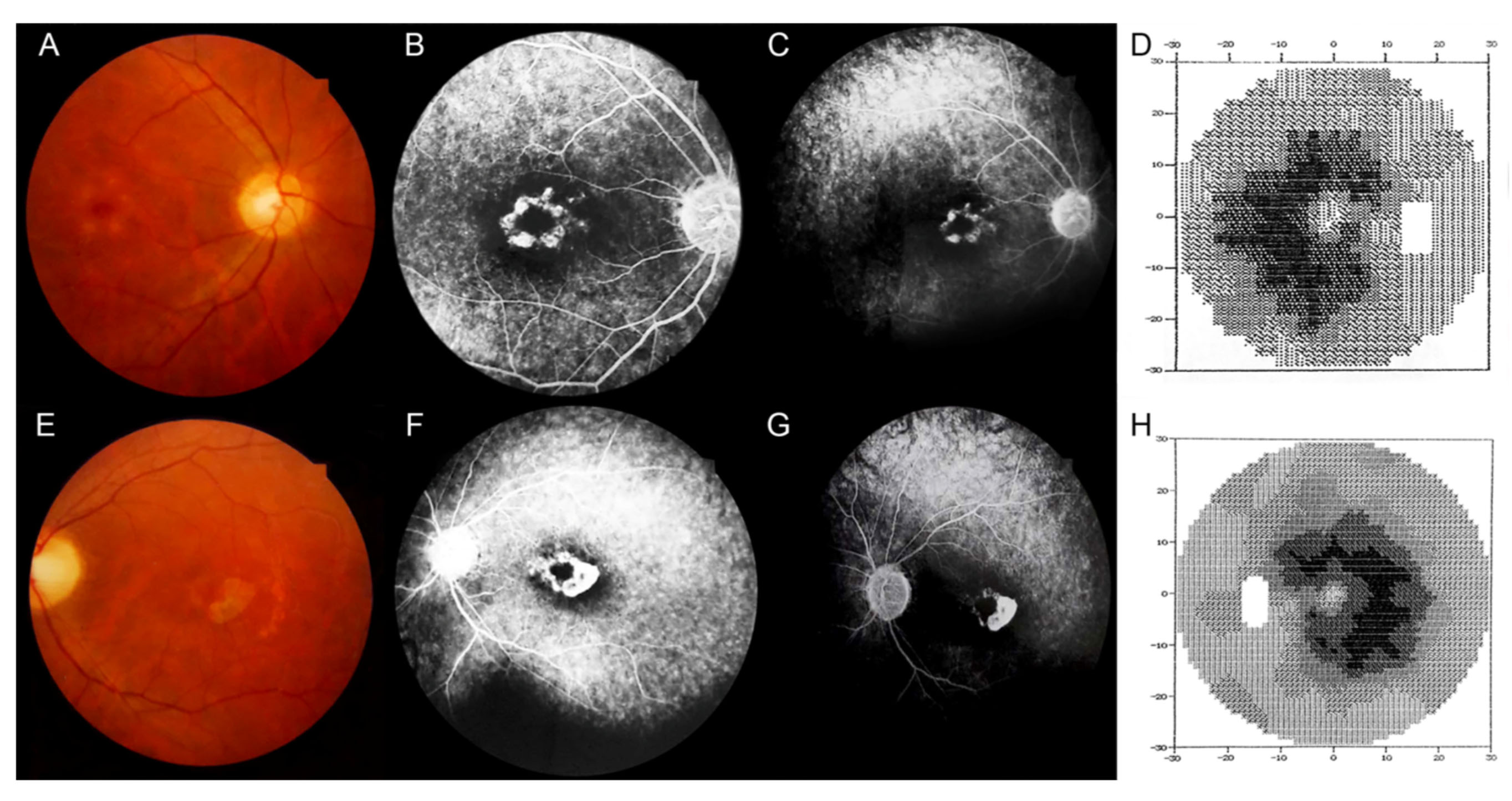
 Figure 2 of
Serra, Mol Vis 2019; 25:814-820.
Figure 2 of
Serra, Mol Vis 2019; 25:814-820.