Figure 6. Expression of GluA2 and PSD-95 on the processes of DACs. Triple immunostaining experiments using antibodies against TH, PSD-95,
and GluA2 (rabbit polyclonal antibodies) were performed in wild-type mouse retinal vertical slices. A single optical section
(0.2 μm thickness) shows colocalization of GluA2 and PSD-95 staining on TH positive processes. A1: DAC processes labeled with TH (red). A2: GluA2 puncta (green). A3: PSD-95 puncta (blue). A4: Overlay. Arrows indicate points of triple-colocalization, showing colocalization of GluA2 and PSD-95 in the DAC processes.
The arrowheads show colocalization of TH and GluA2 in the absence of PSD-95. The open arrowheads point to colocalizations
of TH and PSD-95 in the absence of GluA2. B and C: Overlay images demonstrate colocalizations of TH, PSD-95, and GluA2 staining (arrows). INL, inner nuclear layer; IPL, inner
plexiform layer. Similar results were obtained from three additional mice. Scale bar: 10 μm.
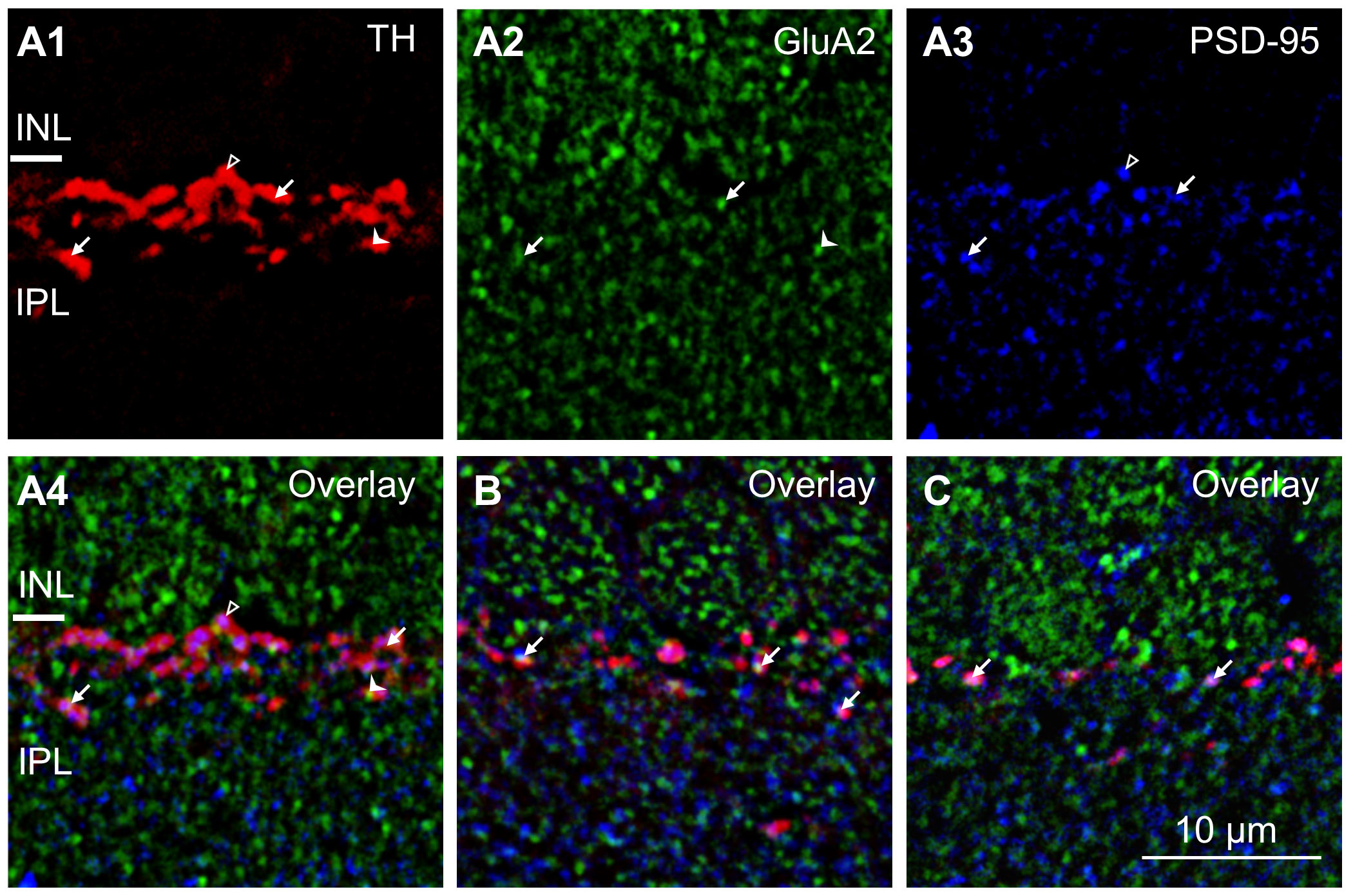
 Figure 6 of
Liu, Mol Vis 2019; 25:780-790.
Figure 6 of
Liu, Mol Vis 2019; 25:780-790.