Figure 5. GluA2 subunits are expressed on TH-positive processes. Double immunostaining experiments using antibodies against TH and GluA2
(a mouse monoclonal antibody) were performed in wild-type mouse retinal vertical slices. A single optical section (0.2 μm
thickness) shows colocalization of GluA2 and TH staining. Left (red), TH-positive cell processes in the IPL. Middle (green),
dense punctate expression of GluA2 in the IPL and sparse expression in the INL. Right, merged image demonstrating putative
colocalization (arrows) of TH and GluA2 on TH-positive processes. INL, inner nuclear layer; IPL, inner plexiform layer. Similar
results were obtained from three additional mice.
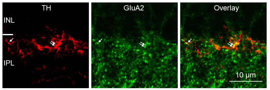
 Figure 5 of
Liu, Mol Vis 2019; 25:780-790.
Figure 5 of
Liu, Mol Vis 2019; 25:780-790.