Figure 1. CECs in MitoM and RestM culture conditions. A:Corneal endothelial cells (CECs) in MitoM culture conditions at P0 before the subculture (10X). B: CECs in MitoM at P1 (10X); and (C) CECs in RestM at P1 (10X). D: CECs in MitoM at P2 (10X); and (E) CECs in RestM at P2 (20X). G: Cellular circularity of human, rabbit basal, MitoM, RestM passage 1 (RestM P1), and RestM passage 2 (RestM P2) CECs. H: Cellular yield analysis of CECs obtained after the first passage in MitoM and RestM.
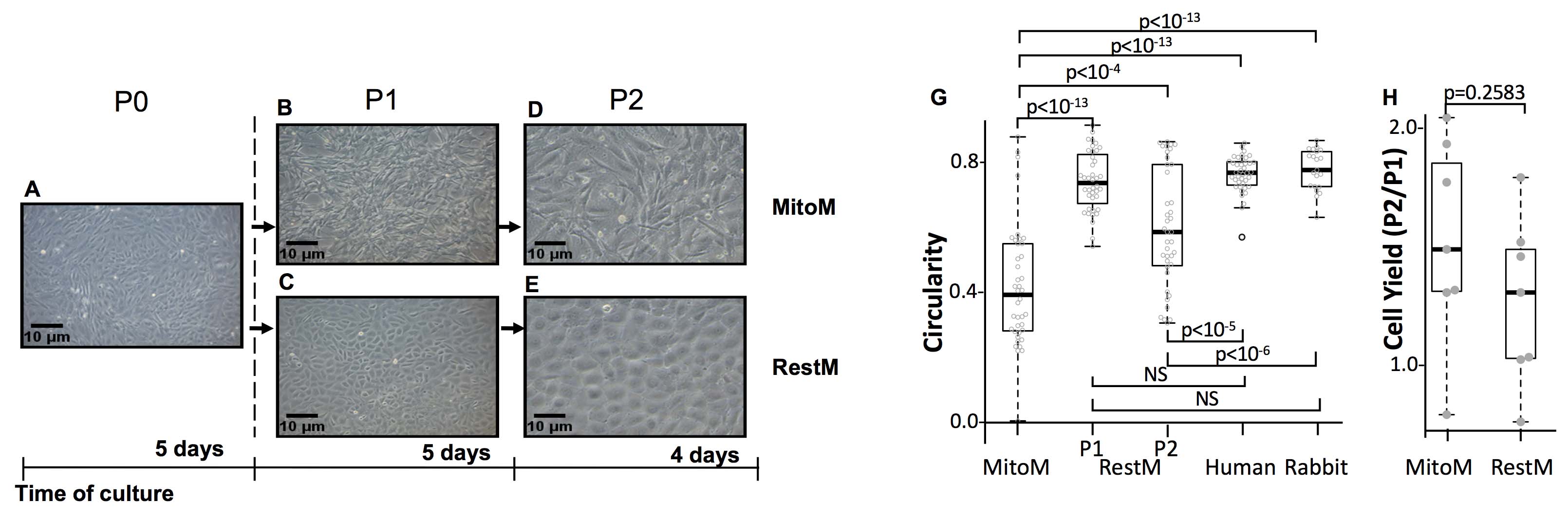
 Figure 1 of
Rodríguez-Barrientos, Mol Vis 2019; 25:745-xxx.
Figure 1 of
Rodríguez-Barrientos, Mol Vis 2019; 25:745-xxx.