Figure 1. The effects of exogenous Sema3A on axon and dendrite differentiation in RGCs. A: Fc-Sema3A (0.5 μg/ml) treatment for 72 h promoted the formation of RGC dendrites and inhibited axon growth, as demonstrated
by immunostaining for neurites and dendrites with III-tubulin and the dendrite marker MAP2 antibodies, respectively. Filled
and empty arrows denote axons and dendrites, respectively. The scale bar is 40 μm. B: The lengths of the longest axon and dendrite per RGC were measured after culture in Fc-Sema3A (0.5 μg/ml) after 72 h (mean±SD;
40-53 cells per treatment). C, D: The numbers of RGCs with different numbers of axons and dendrites were measured following treatment with Fc-Sema3A (1 μg/ml
or 0.5 μg/ml) after 72 h. E: The percentage of bipolar cells was measured following treatment with Fc-Sema3A (1 μg/ml) after 72 h (41-53 cells per treatment).
*p<0.05, **p<0.01.
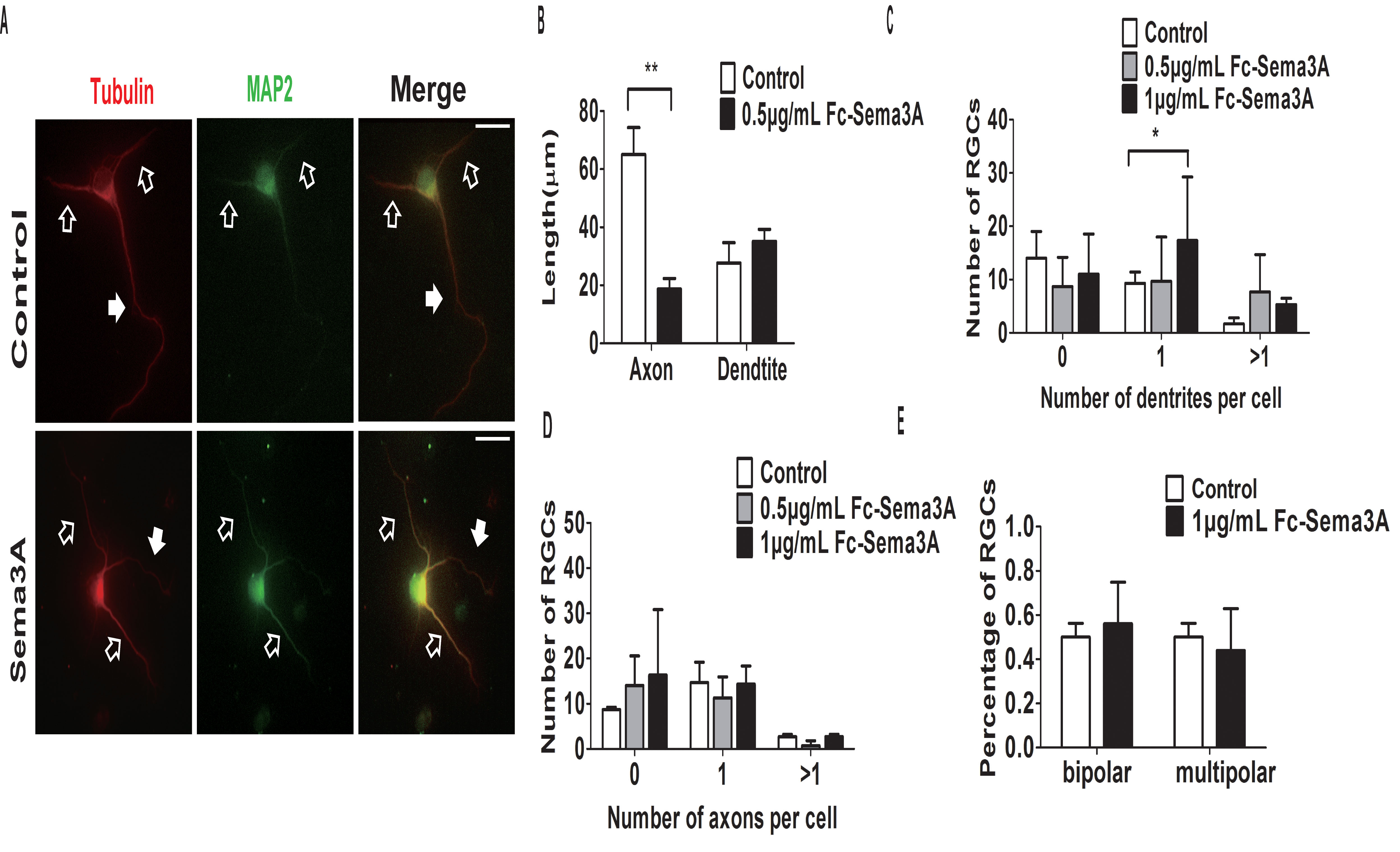
 Figure 1 of
Chan-Juan, Mol Vis 2019; 25:722-730.
Figure 1 of
Chan-Juan, Mol Vis 2019; 25:722-730.