Figure 4. Role of Dp71 in angiogenesis using an aortic ring mouse model. A: Phase contrast photography of typical aortic rings of wild-type (WT; top) and Dp71-null mice (bottom) with neovessels at day 4 (left) and day 7 (right). A visible difference is already present at day 4, and
will increase until day 7 (scale bar=200 µm). B,C: Surface covered by microvessels (in pixels) according to the time (day 3 to 7) in each group in the presence (B) or the absence of vascular endothelial growth factor (VEGF; C). Stars represent statistically significant differences (*p<0.05, **p<0.005, ***p<0.001).
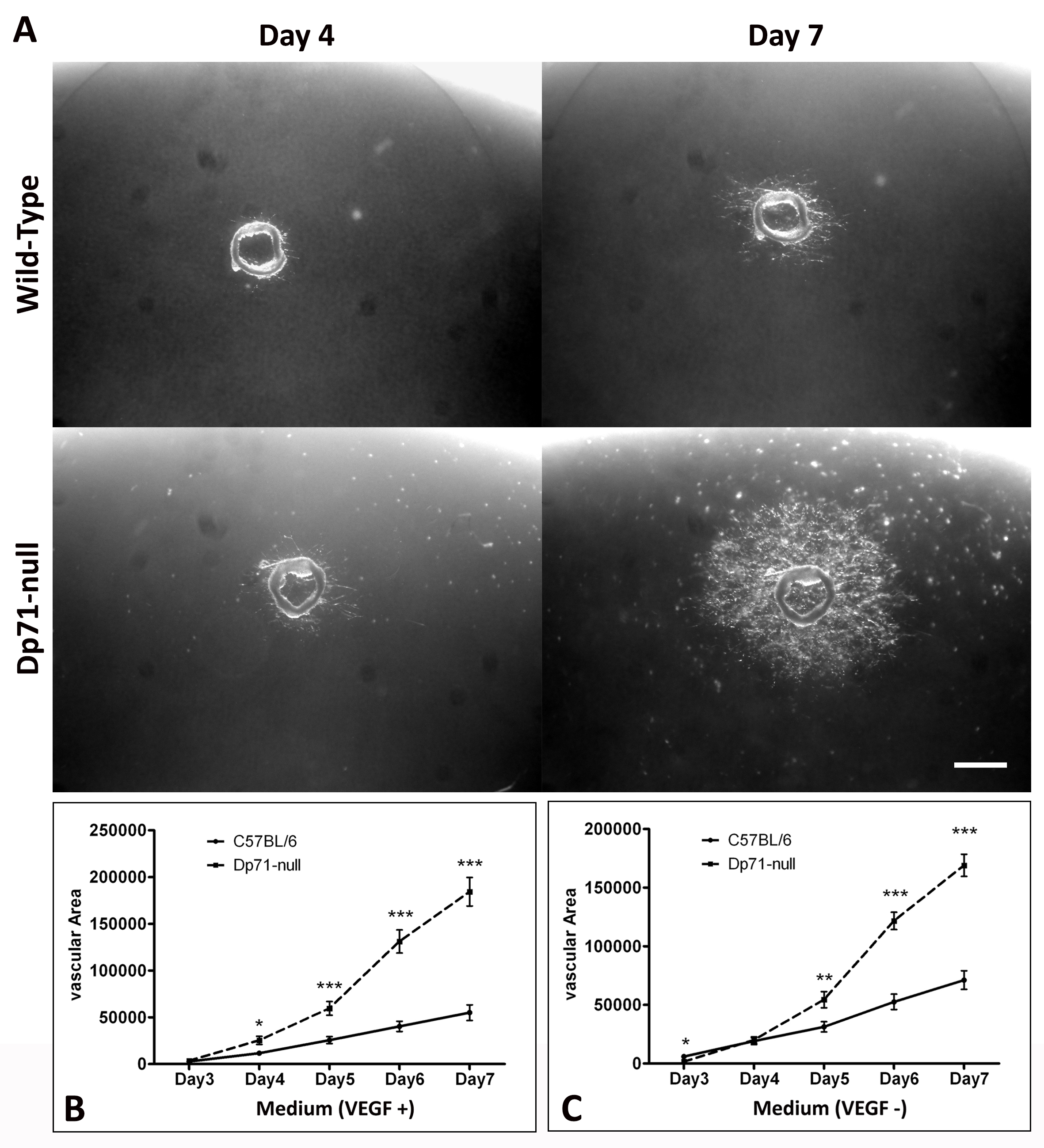
 Figure 4 of
Ortiz, Mol Vis 2019; 25:714-721.
Figure 4 of
Ortiz, Mol Vis 2019; 25:714-721.