Figure 3. Corneal vascularization after or in the absence of injury. A: Visualization of vascular endothelial cells in flatmounted corneas using an anti-CD31 antibody conjugated with fluorescein
isothiocyanate (FITC) of wild-type (WT) or Dp71-null mice, in the absence of injury or 7 days after injury (scale bar=200 µm). B: The corneal vascular area was quantified in WT or Dp71-null mice, in the absence of injury or 7 days after injury. C: The vascular endothelial growth factor (VEGF) concentration was measured with enzyme-linked immunosorbent assay (ELISA)
in WT or Dp71-null mice, in the absence of injury or 7 days after injury.
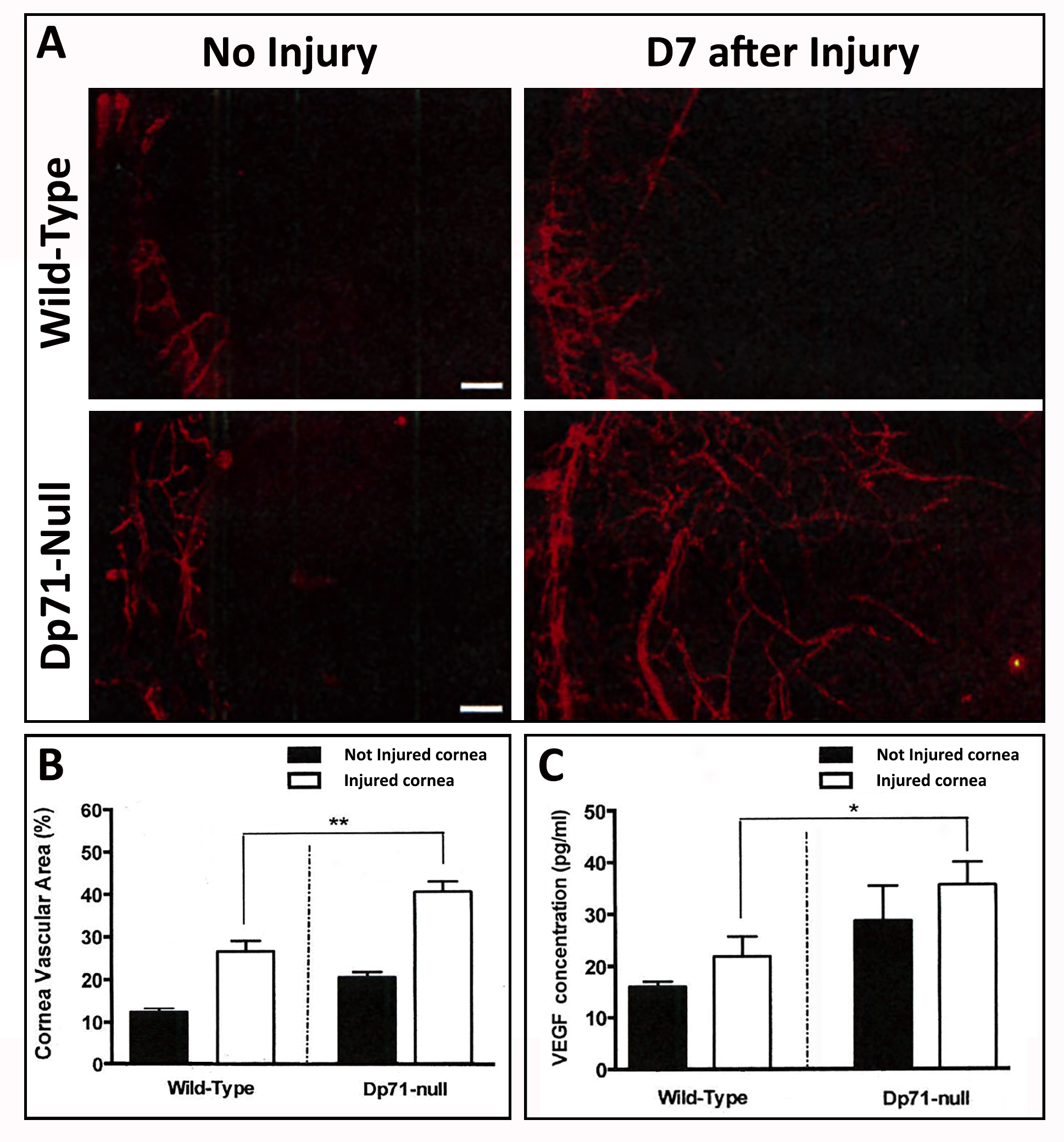
 Figure 3 of
Ortiz, Mol Vis 2019; 25:714-721.
Figure 3 of
Ortiz, Mol Vis 2019; 25:714-721.