Figure 2. Dystrophin expression after or in the absence of corneal injury. A: Immunostaining with a pan-specific antibody directed against dystrophins (H4, green) of the cornea of non-injured wild-type
(WT) and Dp71-null mice (scale bar=20 µm). B: Quantification of H4 staining density with Photoshop software. C: Western blotting of dystrophins on injured and non-injured corneal extracts of WT and Dp71-null mice. D–F: Semiquantification of Dp116 (D), Dp71 (E), and Dp140 (F) protein expression relative to β-actin.
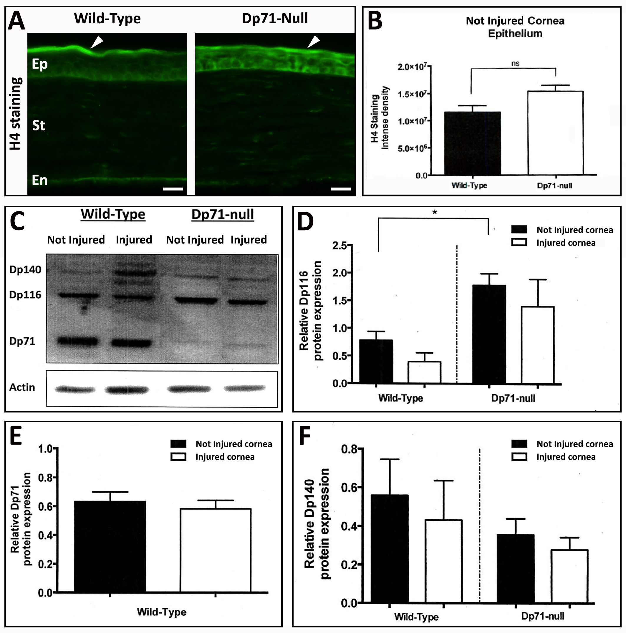
 Figure 2 of
Ortiz, Mol Vis 2019; 25:714-721.
Figure 2 of
Ortiz, Mol Vis 2019; 25:714-721.