Figure 1 of
Ortiz, Mol Vis 2019; 25:714-721.
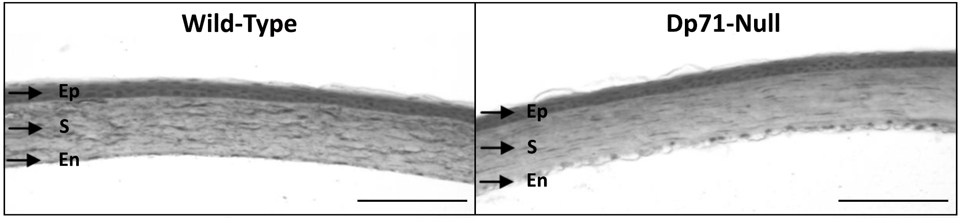
Figure 1.
Corneal histology after hematoxylin-eosin staining of tissue sections of WT and
Dp71
-null mice. Both presented an organized corneal histology with the epithelium (Ep), stroma (S), Descemet membrane, and endothelium (En; scale bar=200 µm).
 Figure 1 of
Ortiz, Mol Vis 2019; 25:714-721.
Figure 1 of
Ortiz, Mol Vis 2019; 25:714-721.