Figure 6. The Sox9 knockdown decreased the expression of GFAP as measured by immunohistochemistry. In both the control and Sox9-shRNA
groups, the labeling intensity of GFAP increased, and the spatial distribution expanded on days 3, 7, and 14 after light expose.
However, the GFAP staining intensity and spatial distribution in the Sox9-shRNA group were obviously attenuated compared with
the control at each time point. All photomicrographs were taken under a photomicroscope within identical parameters. Scale
bar, 50 μm.
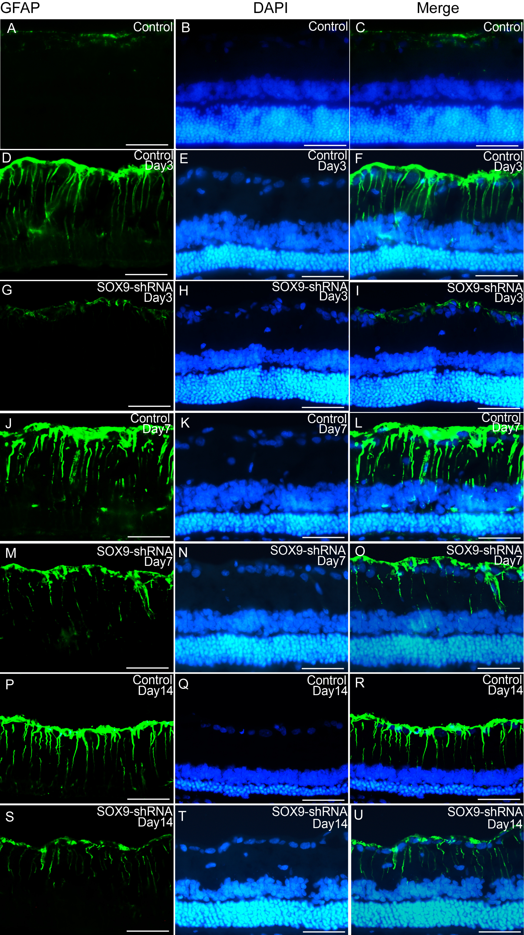
 Figure 6 of
Wang, Mol Vis 2019; 25:703-713.
Figure 6 of
Wang, Mol Vis 2019; 25:703-713.