Figure 1. Validation of the Sox9 knockdown status of the Sox9-shRNA lentiviral vector after two weeks of intravitreal injection using
co-labeling immunostaining and western blotting. A: Sox9 was mainly located in the Müller cell nucleus in both the control group and the Sox9-shRNA group and there was relatively
weaker staining of Sox9 in the Sox9-shRNA group compared with the control group. Scale bar, 50 μm. B: Western blot analysis of the Sox9 protein in the retinas from the control and Sox9-shRNA groups. Β-actin is the loading
control. Relative expression of the Sox9 protein in the Sox9-shRNA group is significantly reduced compared with the control
at every studied time point. Data are expressed as a percentage of the control values presented as mean ± SD (n = 3), t test,
*p<0.05.
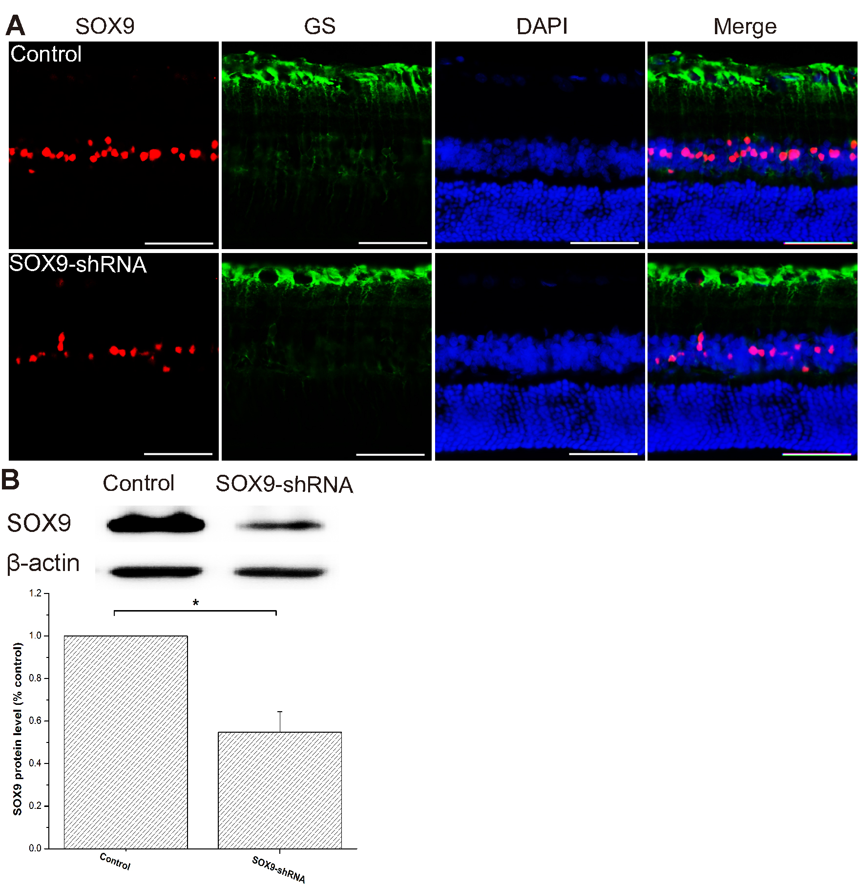
 Figure 1 of
Wang, Mol Vis 2019; 25:703-713.
Figure 1 of
Wang, Mol Vis 2019; 25:703-713.