Figure 4. Schematic representation of retinal pigment epithelium (RPE) loss and repair. A: Small druse (<63 µm). The RPE pattern of a rosette filling in a defect is consistent with apoptosis of a single RPE cell
overlying the small druse. B: Intermediate drusen (63-124 µm). The RPE pattern of complete loss overlying the intermediate druse is consistent with an
autophagy mechanism of RPE cell loss. C: Large drusen (≥125 µm). The pattern of RPE loss with remaining basal cytoplasm overlying large drusen is consistent with
necroptosis as a mechanism. D: Basal laminar deposit. RPE loss in sheets with preservation of basal cytoplasm and later multi-nucleated RPE is consistent
with necroptosis, cell migration, and fusion as a mechanism of repair.
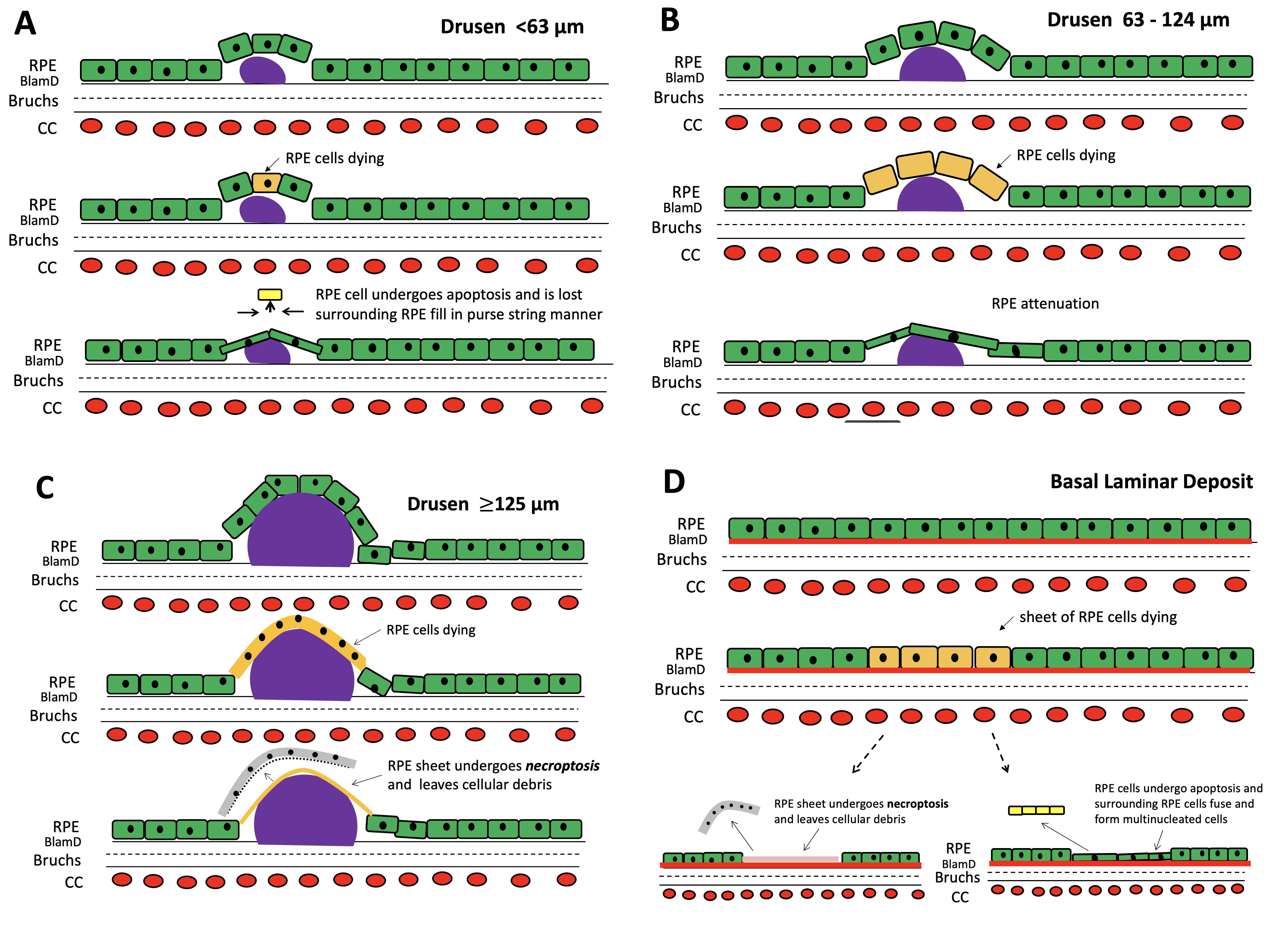
 Figure 4 of
Zhang, Mol Vis 2019; 25:70-78.
Figure 4 of
Zhang, Mol Vis 2019; 25:70-78.