Figure 3. Drusen types, basal laminar deposit (BlamD), and corresponding retinal pigment epithelium (RPE) morphology. A-D. Small drusen
(<63 µm). A and B: Small drusen are present between the RPE (arrowhead) and Bruch's membrane (arrow). C: The drusen are ultrastructurally composed of heterogeneous, variable electron-dense material (arrow). The RPE (R) overlying
the drusen is intact (5,800X). D: The corresponding RPE flatmount shows a rosette overlying the small drusen (arrow) autofluorescence are shown in blue. E-H: Intermediate drusen (63-124 µm). E and F: The drusen form mounds between the RPE (arrowhead) basal lamina and inner collagenous layer of the Bruch’s membrane (arrow);
choroid (C). There is RPE loss overlying the apex of the druse. G. Intermediate druse is composed of heterogeneous electron dense material
(arrow), which was not covered by RPE (4,800X). H. The corresponding RPE flatmount shows an irregular RPE surrounding the
druse and RPE loss overlying the druse (arrow). I-L: Large drusen. I and J: The RPE overlying the large drusen shows loss of apical cytoplasm; the basal cytoplasm remains (arrowhead; 100X); choroid
(C); Bruch’s membrane (arrow), K: The drusen are ultrastructurally composed of more homogenous material (arrow) than small and intermediate drusen. The basal
cytoplasm of the RPE (R) overlying the drusen is intact (5800X). L: The corresponding RPE flatmount loss of RPE cell borders and apparent fusion of RPE (green) with multiple nuclei (red; arrows)
adjacent to the large druse (arrowhead). M-P: Basal laminar deposit (BlamD). M and N: The RPE/BlamD complex is detached (*) from the underlying Bruch’s membrane and choroid. The RPE is intact. O: The BlamD (between arrows) is interposed between the plasma membrane and basal lamina of the RPE; choroid (C); RPE (arrowhead);
Bruch’s membrane (arrow) (5,800X) P: The corresponding RPE (green), including rosette formation (arrowhead), marked changes in cell size and shape, and RPE loss
in areas (arrow). E,I (hematoxylin, high power objective [HPO]) M (periodic acid-Schiff, HPO), B, F, J, N (toluidine blue, HPO), C and K (osmium tetroxide/lead citrate, 5,800X), G and O (osmium tetroxide/lead citrate, 4,800X), D, H, L, P (AF635-phalloidin/propidium iodide 100X) RPE are green, druse and nucleus are red and autofluorescence are blue.
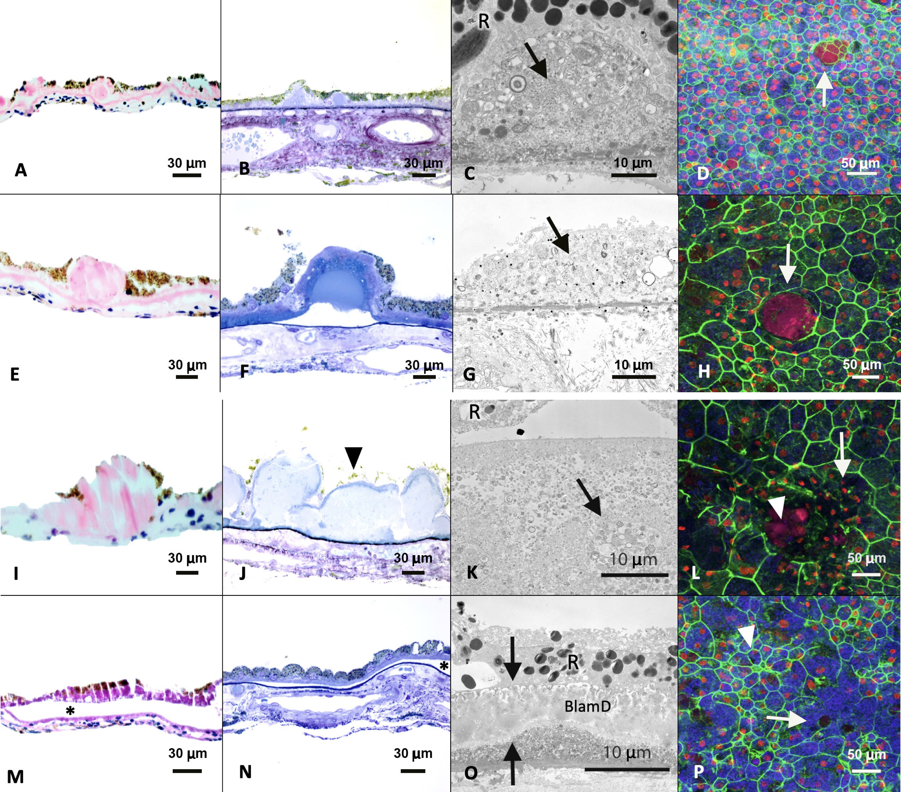
 Figure 3 of
Zhang, Mol Vis 2019; 25:70-78.
Figure 3 of
Zhang, Mol Vis 2019; 25:70-78.