Figure 2. Representative images of retinal pigment epithelium (RPE) flatmounts. A1,2: Gross inspection of the posterior preparation of eyes in patient 1 with age-related macular degeneration (AMD) showed
pigment mottling and drusen (arrows) within the macular area; optic nerve (asterix). A3,4: The RPE/Bruch's membrane/choroid preparations with preservation of the pigment mottling and drusen (arrowhead). B: RPE flatmount preparation showed normal RPE changes due to aging from a donor without AMD. C: RPE flatmount preparation showed drusen (arrow) and RPE atrophy (arrowhead) in right eye of patient 1 with AMD. D: RPE flatmount preparation showed drusen (arrow) and RPE atrophy (arrowhead) in left eye of patient 1 with AMD. The reconstructed
surface area measured about 4 x 4 mm. (Panels C and D, RPE border is green, nucleus is red).
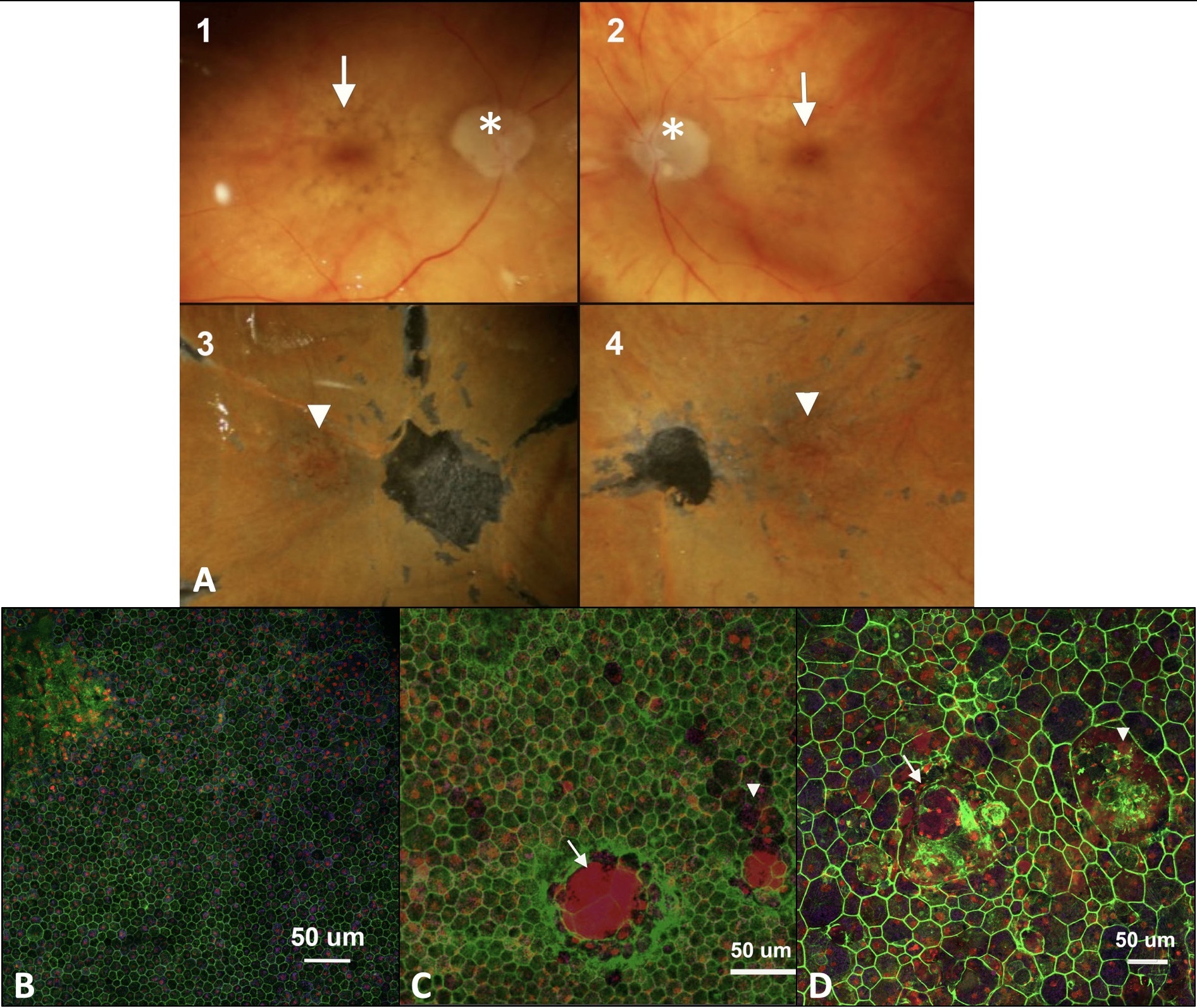
 Figure 2 of
Zhang, Mol Vis 2019; 25:70-78.
Figure 2 of
Zhang, Mol Vis 2019; 25:70-78.