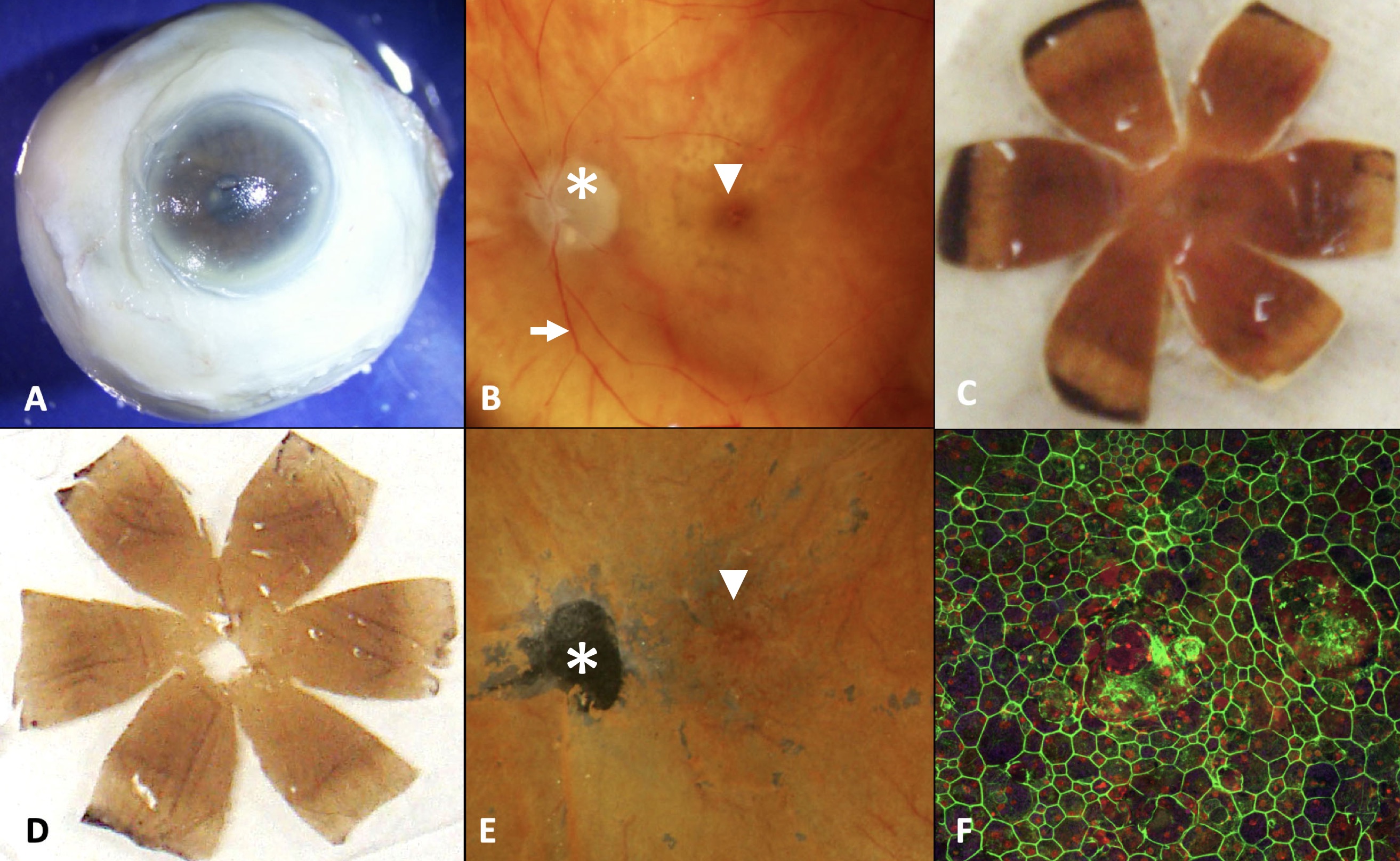
Figure 1. Retinal pigment epithelium (RPE) flatmount preparation.
A: Post-mortem eye is grossly inspected.
B: The eye is opened coronally and the anterior portion of the eye is removed, thus leaving a posterior optic nerve (asterix),
sclera, choroid, RPE, retina including the macula (arrowhead) with the retinal vessels (arrow) and vitreous preparation.
C: The posterior preparation is microdissected into six radial anterior-posterior petals.
D: The sclera is removed from the petals leaving a retina, RPE, choroid preparation.
E: The retina and vitreous are removed, thus leaving an RPE, Bruch's membrane, choroid preparation; macula (arrowhead); optic
nerve defect (asterix).
F: The RPE was stained with AF635-phalloidin pseudo colored with green and examined by confocal microscopy, thus yielding an
RPE mosaic. This method of preparation is similar to that which we previously reported [
13].
 Figure 1 of
Zhang, Mol Vis 2019; 25:70-78.
Figure 1 of
Zhang, Mol Vis 2019; 25:70-78.