Figure 3. Fundus autofluorescence (FAF) images. A: Multiple hypo-autofluorescence dots without a bull’s-eye pattern in patient H27. Band E: Typical FAF images of STGD, with round macular hypo-autofluorescence and multiple dots (H75 and H278). C: Hypo-autofluorescence on the entire macula in a patient with CRD (H144). D: Bull’s-eye maculopathy without flecks (H234). F: FAF images in a patient with fundus flavimaculatus (H830).
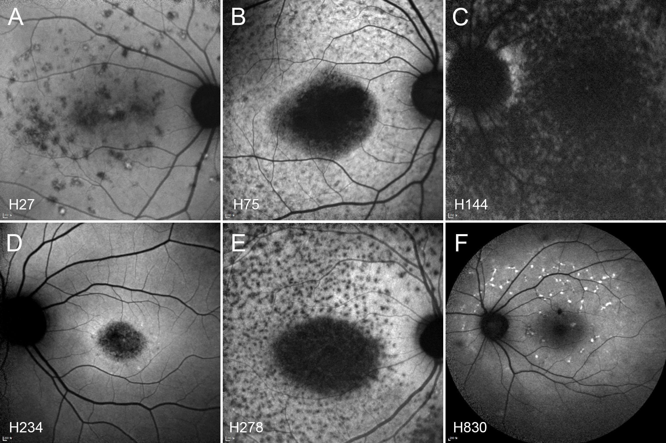
 Figure 3 of
Joo, Mol Vis 2019; 25:679-690.
Figure 3 of
Joo, Mol Vis 2019; 25:679-690.