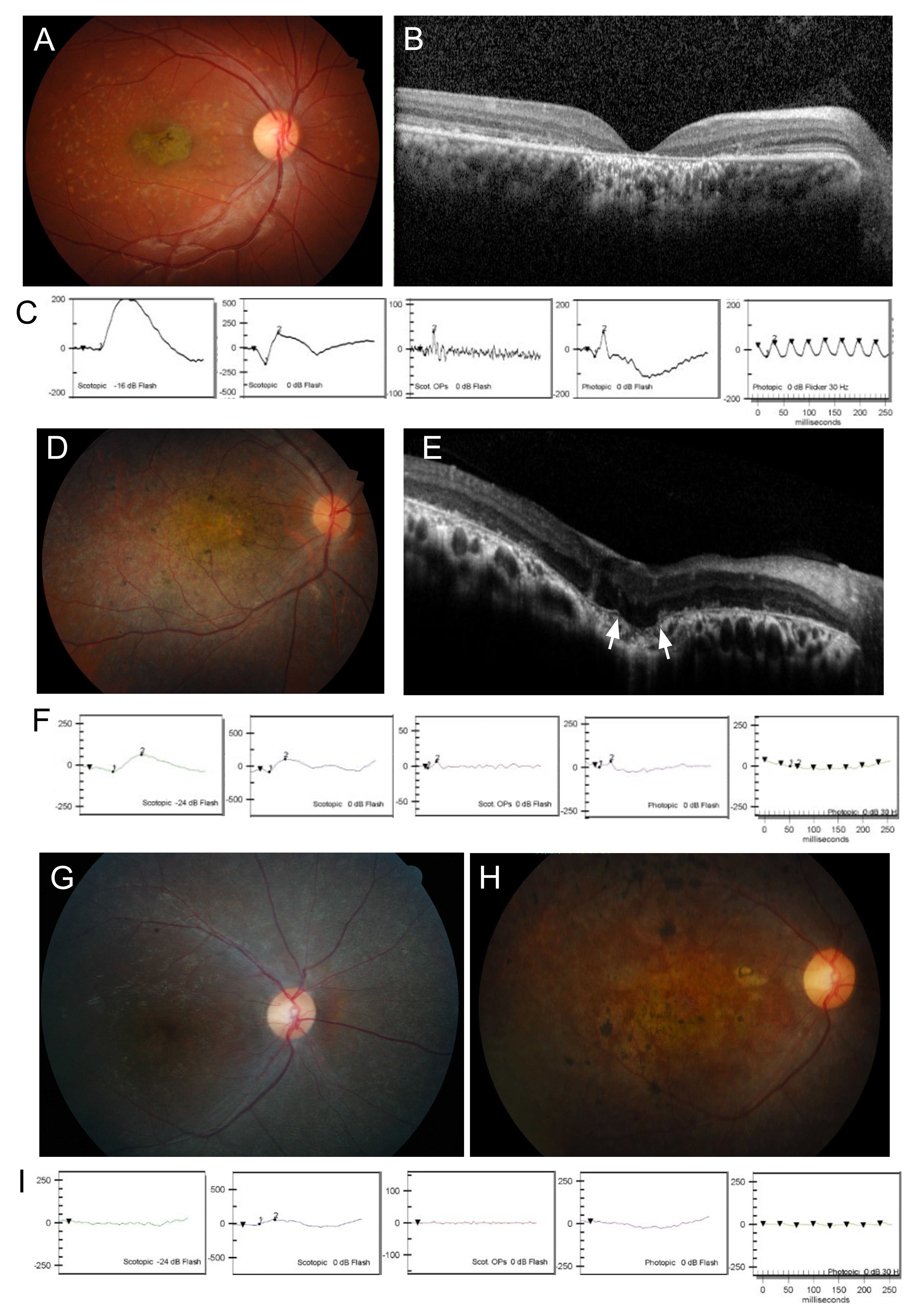
Figure 2. Clinical features of ABCA4-associated retinopathies. A, D, G, and H: Fundus photography. BandE: Optical coherence tomography (OCT). C,F, and I: Electroretinograms (ERGs). A, B, andC: The ophthalmologic examination of patient H147 showed typical features of STDG. Atrophy of photoreceptors and RPE in the
macula is shown on fundus photography and OCT. D, E, and F: Patient H145 showed CRD with macular degeneration and reduced cone response. The white arrows indicate choroidal excavation
and defect of the Bruch’s membrane. G and H: Fundus examination of patient H91 at 10 and 19 years of age, respectively. I: Both the cone and the rod responses on the
ERG at the age of 10 years were strikingly decreased, suggesting typical features of RP.

 Figure 2 of
Joo, Mol Vis 2019; 25:679-690.
  Figure 2 of
Joo, Mol Vis 2019; 25:679-690.