Figure 5. 9CRAL-expedited photoreceptor development. A: Representative images of immunostained sections of H9 retinal organoids supplemented with either all-trans retinoic acid
(ATRA; left) or 9-cis retinal (9CRAL; right). D90 (top) 10 μm sections were immunolabeled for pan-photoreceptor marker recoverin
(green) and retinal progenitor cell marker CHX10 (red). D120 sections (bottom) were immunolabeled for rod photoreceptor marker
rhodopsin (green), cone photoreceptor marker OPN1SW (red), and L/M cone photoreceptor marker OPN1L/MW (magenta). Nuclei were
stained with 4′,6-diamidino-2-phenylindole (DAPI, blue). Arrowheads indicate relevant staining of a specific marker. Scale
bar, 10 μm. B: Immunoblot showing rhodopsin expression in H9 ATRA and 9CRAL organoids at D120 (individual replicates are shown). The asterisk
denotes a second band which is likely to be dimerization of rhodopsin. γ-tubulin is included as the protein loading control
for protein amounts. C: Transmission electron microscopy of longitudinal sections at D130, showing inner segments and cilia. Hollow, solid, and
v-shaped arrowheads indicate the relevant structure of the photoreceptor cilium, mitochondria, and inner segments, respectively.
Magnifications (scale bars) in the right panel are the same as shown in the left.
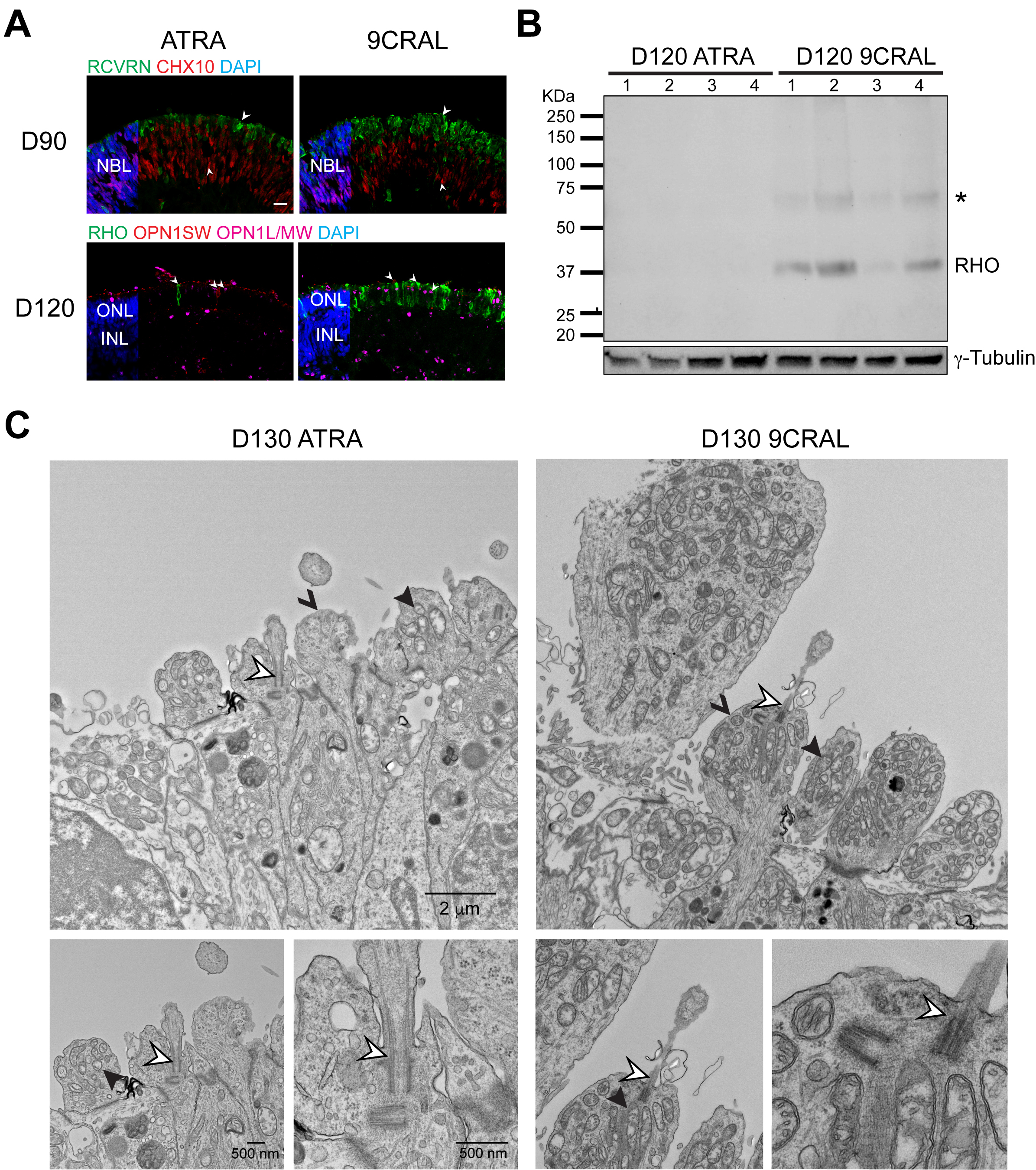
 Figure 5 of
Kaya, Mol Vis 2019; 25:663-678.
Figure 5 of
Kaya, Mol Vis 2019; 25:663-678.