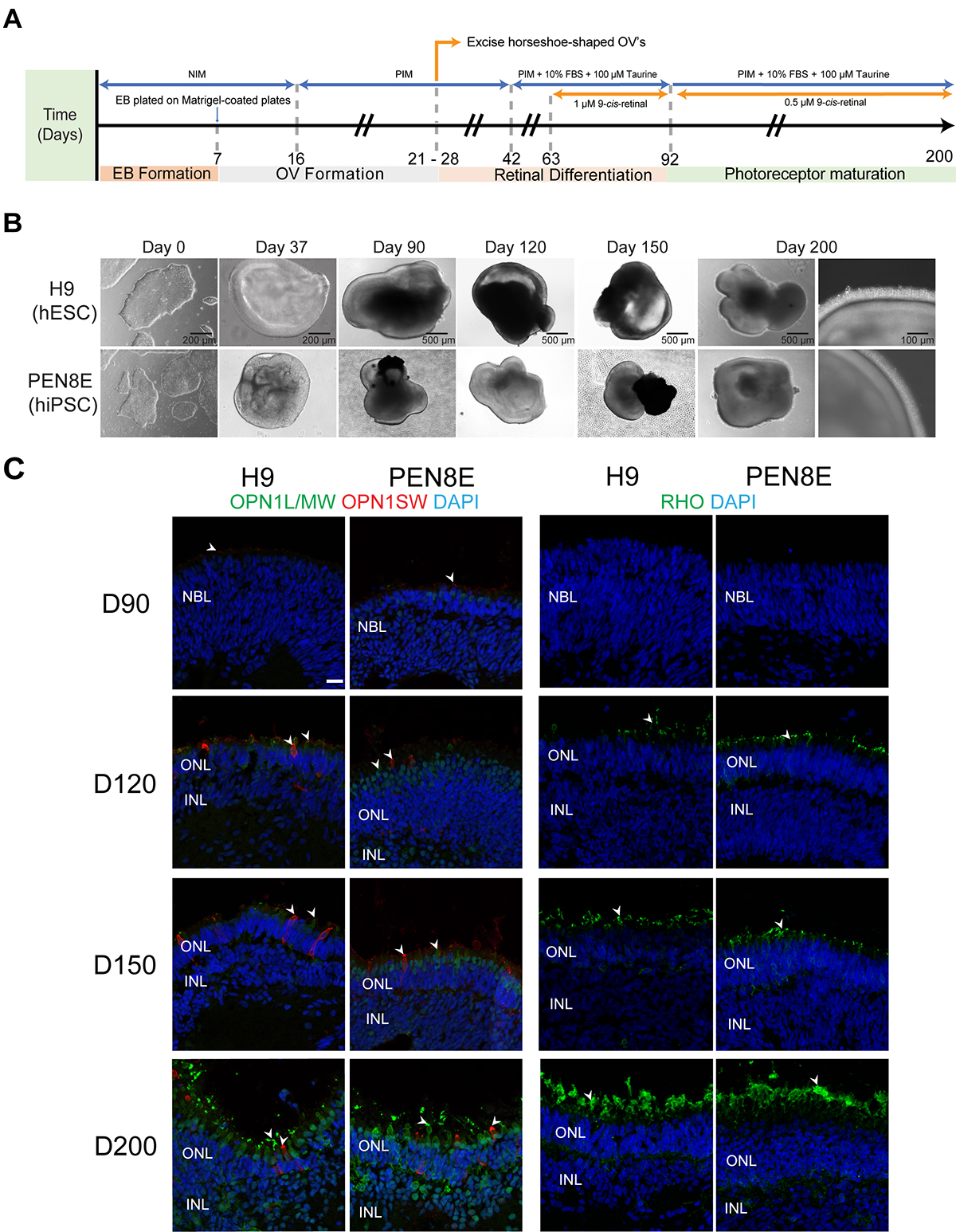
Figure 1. Differentiation of human pluripotent stem cells into retinal organoids.
A: Differentiation protocol used in this study, modified from [
29,
33]. Numbers under the arrow indicate the differentiation day. NIM: neural induction medium; PIM: photoreceptor induction medium;
EB: embryoid body; OV: optic vesicle; FBS: fetal bovine serum.
B: Representative bright-field images of human embryonic stem cells (hESCs; H9) and human induced pluripotent stem cells (hiPSCs;
PEN8E), and of differentiating organoids (from D37 to D200).
C: Immunohistochemistry analysis of H9- and PEN8E-derived retinal organoids using marker antibodies for cones (OPN1L/MW, OPN1SW)
and rods (RHO). Nuclei were stained with 4′,6-diamidino-2-phenylindole (DAPI, blue). Arrowheads indicate relevant staining
of each marker. Scale bar: 20 μm. D: differentiation day; NBL: neuroblastic layer; ONL: outer nuclear layer; INL, inner nuclear
layer; OPL: outer plexiform layer; IPL: inner plexiform layer.
 Figure 1 of
Kaya, Mol Vis 2019; 25:663-678.
Figure 1 of
Kaya, Mol Vis 2019; 25:663-678.