Figure 1. Multimodal imaging testing of three patients with the Hom.c.802–8_810del17insGC mutation. Patient 4: Color fundus images (A) show normal vasculature, RPE atrophy, choroidal sclerosis, and a rare distribution of crystallization. Fundus fluorescein
angiography (FFA) (B) shows atrophy of RPE and choroidal capillaries with a mottled hypofluorescence. Visual field (C) analysis indicates decreased visual acuity (VA) and irregular visual field defects. Spectral domain optical coherence tomography
(SD-OCT) (D and E) shows the disappearance of the inner segments/outer segments (IS/OS) band, ellipsoid zone (EZ), outer nuclear layer, and
outer limiting membrane, and thinning of the thickness in the RPE, retina, and choroid. Patient 6: Color fundus images (F) show diffused glistening yellow crystal deposits in the posterior pole and atrophic changes in the RPE. The retinal vessels
appear normal. FFA (G) shows diffused spiced-salt-shaped hyperfluorescence in the posterior pole. Visual field changes (H) indicate paracentral absolute scotoma and decreased VA at 30°. SD-OCT (I,J) show the disappearance of the IS/OS band and the EZ. The thickness of the RPE, retina, and choroid indicates thinning. Patient 7: Color fundus images (K) show sparkling yellowish-white fine spots in the fundus and a submacular hemorrhage in the left eye. FFA (L) shows spiced-salt-shaped and map-shaped hypoautofluorescence. The visual field (M) shows mild reduced VA at 30°. SD-OCT (N, O) shows that the structure of the outer retina is complete, with only thinning of the choroid. Note A limited bulge is observed
below the foveal in the left eye.
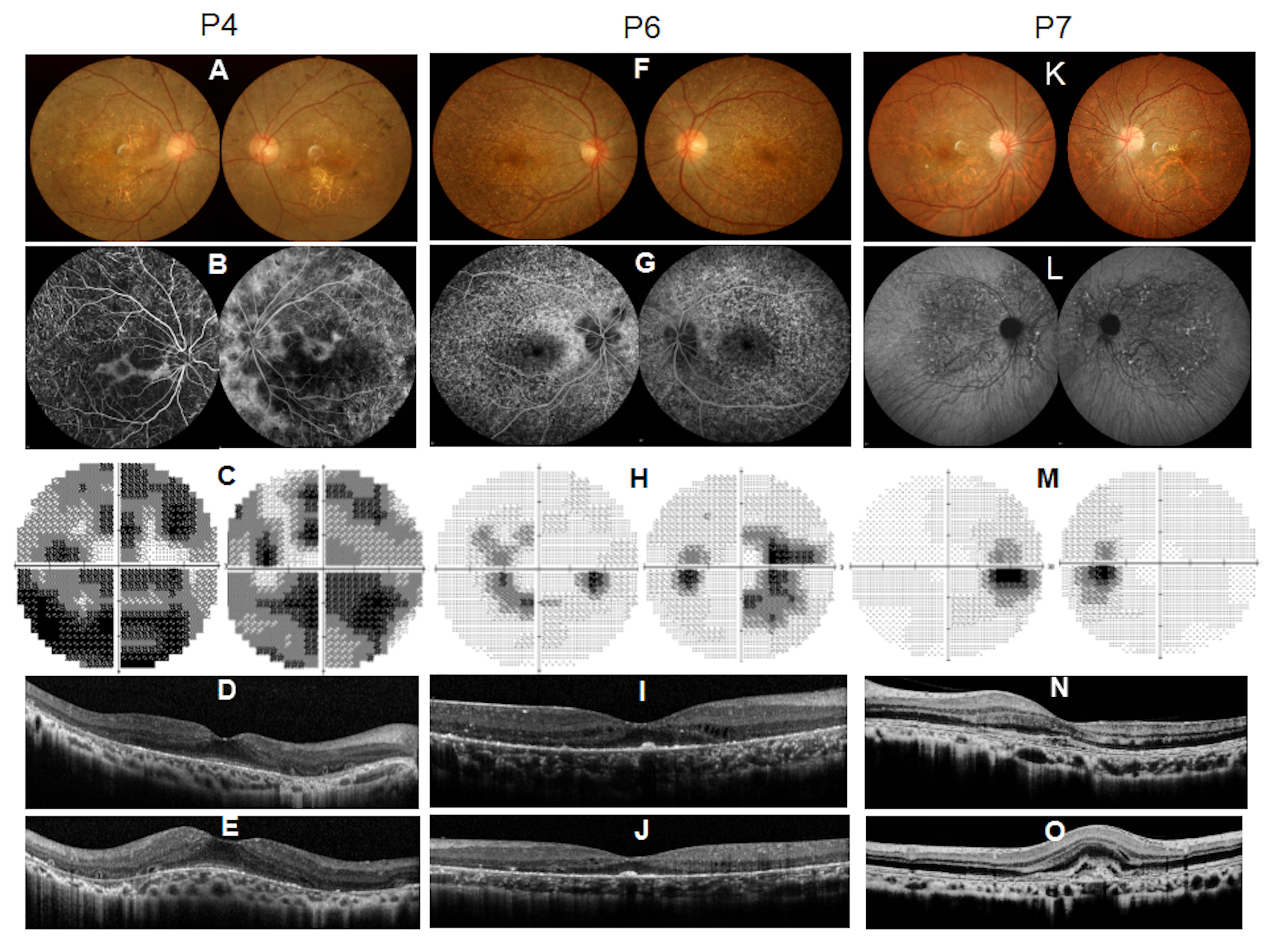
 Figure 1 of
Meng, Mol Vis 2019; 25:654-662.
Figure 1 of
Meng, Mol Vis 2019; 25:654-662.