Figure 6. Completely blocking the BMP pathway inhibited redifferentiation in low passage fetal RPE cells and upregulated GREM1A: Four groups: the control group, SB group, S+L group, and LDN group. After treatment, the phase contrast micrographs demonstrate
that the cells treated with dual pathway inhibitors or LDN193189 had poor differentiation, but other groups without BMP inhibitors
grew well and are pigmented. B: The quantitative PCR (qPCR) results show that the expression of RPE function-specific genes in the S+L group and LDN group
cells is much lower than cells in other groups, but the control group cells and the SB group cells had no statistically significant
differences. C: The expression of GREM1 in the S+L group and LDN group cells is upregulated compared to that of the other groups without the BMP inhibitor treatment.
Scale bar: Ph: 100 μm; Bf: 500 μm. Data are shown as the mean ± standard error of mean (SEM), n=3, **p<0.01 versus P2 control
group. Ph: Phase contrast; Bf: Bright-field; SB group: SB431542 group; S+L group: SB431542+LDN193189 group; LDN group: LDN193189
group.
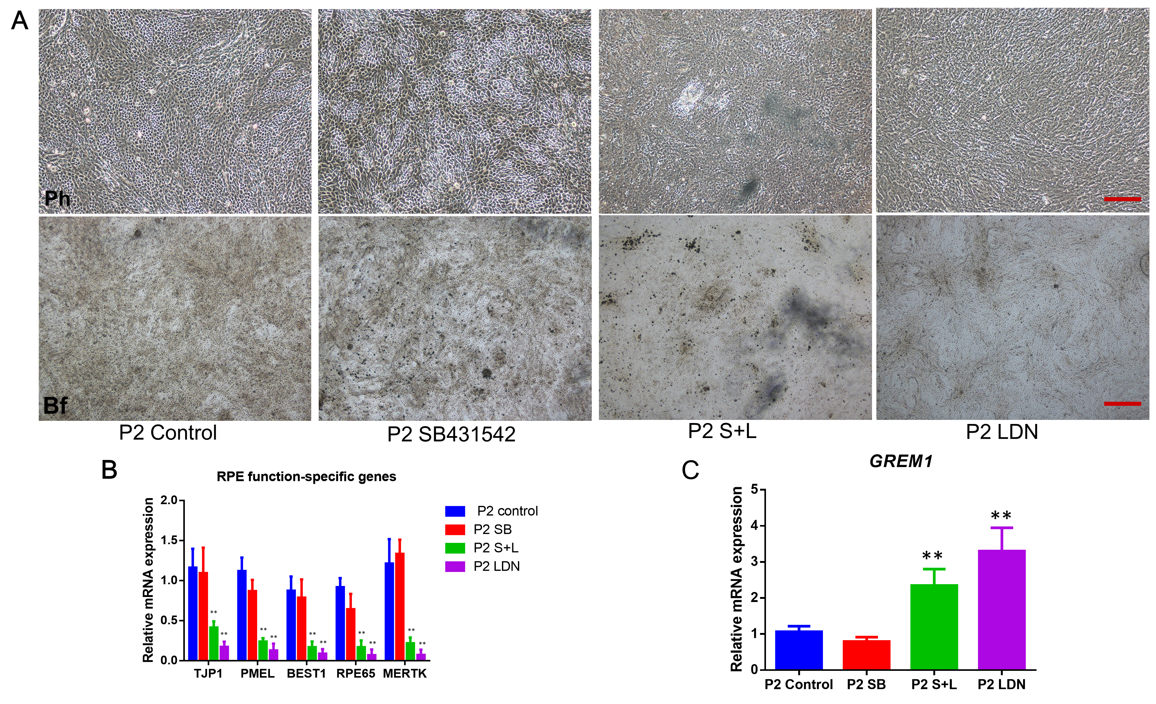
 Figure 6 of
Li, Mol Vis 2019; 25:625-635.
Figure 6 of
Li, Mol Vis 2019; 25:625-635.