Figure 4. Exogenous Gremlin-1 induced EMT by upregulating SNAI1 and downregulating the expression of key transcription factors in fetal RPE cells. A: The phase contrast micrographs show that passage 2 cells treated with recombinant human Gremlin-1 develop a mesenchymal-like
appearance compared with the control group cells. B: Cell scratch wound assays show that the cells with the Gremlin-1 treatment have more cell migration after 2 days. C: The quantitative PCR (qPCR) results show that the EMT-related genes, SNAI1 and VIM, are upregulated after Gremlin-1 treatment, while the RPE-related genes (MITF, OTX2, and RPE65) are downregulated. D: The western blotting results show that Gremlin-1 reduces ZO-1 expression and increases vimentin, and after Gremlin-1 treatment,
p-Smad2 and Smad4 are upregulated. Actin serves as an internal reference in the western blots. Scale bar: Ph: 100 μm. Data
are shown as the mean ± standard error of mean (SEM), n=3, ***p<0.001 versus control. Ph: Phase contrast; p-Smad2: phosphorylated-Smad2.
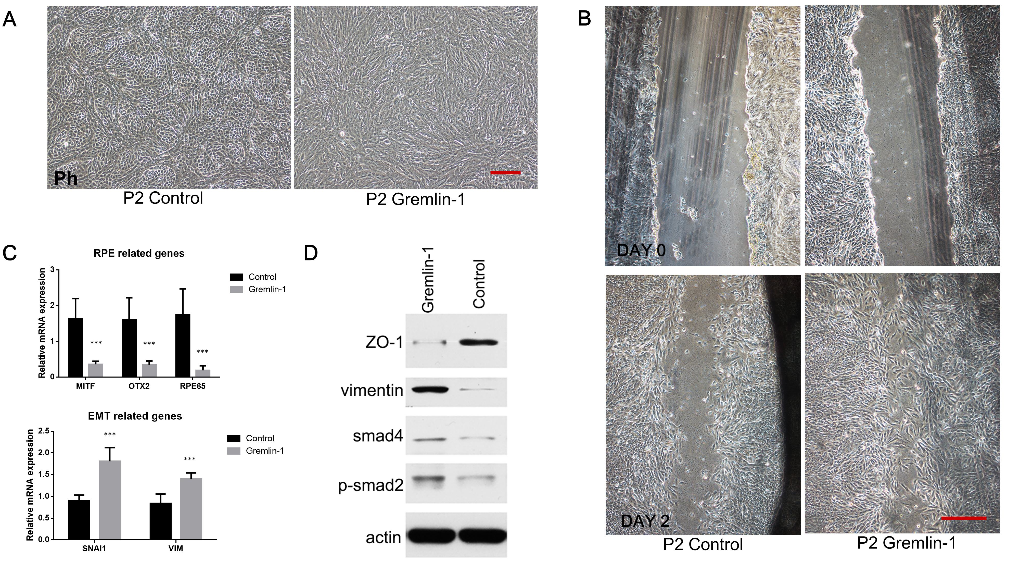
 Figure 4 of
Li, Mol Vis 2019; 25:625-635.
Figure 4 of
Li, Mol Vis 2019; 25:625-635.