Figure 2. The expression of GREM1 was upregulated in the epithelial-mesenchymal transition of fetal RPE cells. A: The phase contrast and bright-field micrographs show that fetal RPE cells treated with SB431542 every day maintained a cobblestone-like
appearance and pigments until passage 11, and then they developed a fibroblast-like appearance and lost their pigment. B: When cells are repetitively passaged, the expression of RPE function-specific genes shows a relatively declining trend,
and passage 11 cells have the lowest expression levels. C: The expression of GREM1 is increased gradually with repetitive passages, and passage 11 cells have the highest expression compared to the other passages.
Scale bar: Ph: 100 μm, Bf: 500 μm. Data are shown as the mean ± standard error of mean (SEM), n=3, **p<0.0001, *p<0.001, #p<0.05
versus P2 SB431542. Ph: Phase contrast; Bf: Bright-field SB: SB431542.
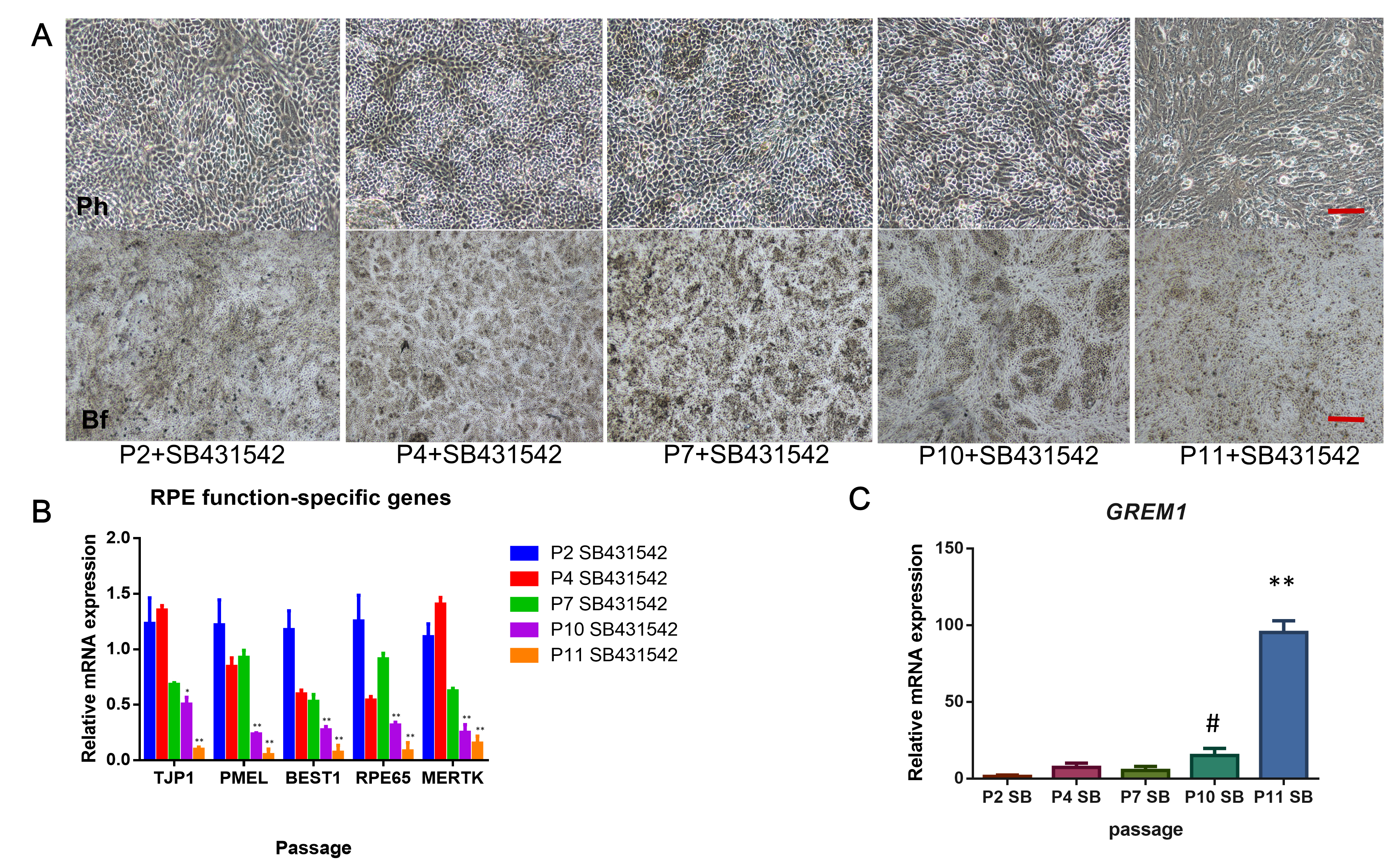
 Figure 2 of
Li, Mol Vis 2019; 25:625-635.
Figure 2 of
Li, Mol Vis 2019; 25:625-635.