Figure 1. The expression of GREM1 was upregulated in the epithelial-mesenchymal transition of fetal RPE cells. A: On day 32, passage 2 cells cultured without any treatment have a normal cobblestone-like appearance in phase contrast micrographs
and more pigment in the bright-field micrographs, but passage 4 cells have a fibroblast-like shape and lost pigment. B: The related expression of RPE function-specific genes, such as TJP1, PMEL, BEST1, RPE65, and MERTK, decreased in passage 4 cells compared to passage cells. C: In passage 2 cells, CDH1 had higher expression compared to passage 4 cells, and the western blotting results show that P-cadherin, which is a specific
epithelial marker in RPE, is expressed more in passage 2 cells. D: VIM, which is a marker of epithelium-mesenchymal transition (EMT), has lower expression than passage 4 cells. E: The related expression of GREM1 in passage 2 cells is lower than that of passage 4 cells. Scale bars: Ph: 100 μm, Bf: 500 μm. Data are shown as the mean
± standard error of mean (SEM), n=3, ****p<0.0001, ***p<0.001 versus P2 blank; ####p<0.0001, #p<0.05 versus P4 blank; Ph:
Phase contrast; Bf: Bright-field CDH1: epithelial marker; VIM: EMT marker.
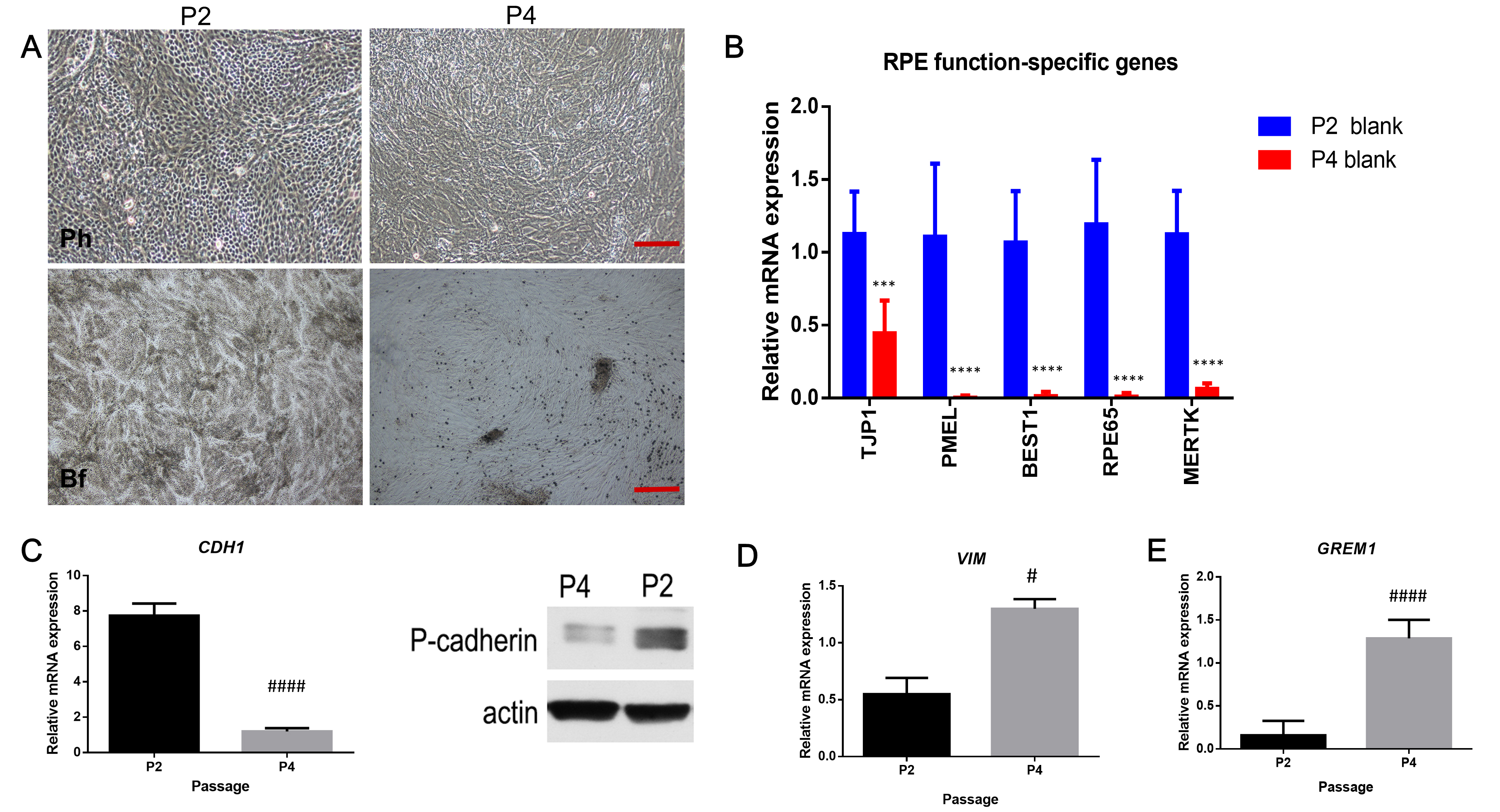
 Figure 1 of
Li, Mol Vis 2019; 25:625-635.
Figure 1 of
Li, Mol Vis 2019; 25:625-635.