Figure 5. Fundus and angiographic images of family D with familiar exudative vitreoretinopathy. A, B: Fundus photographs of the proband show fibrovascular tissue with retinal folds involving the macula in two eyes. C, D: For his affected father, angiography reveals aberrant vessels and avascular area in the peripheral retina. E, F: His mother has normal retinal vasculature demonstrated with angiography.
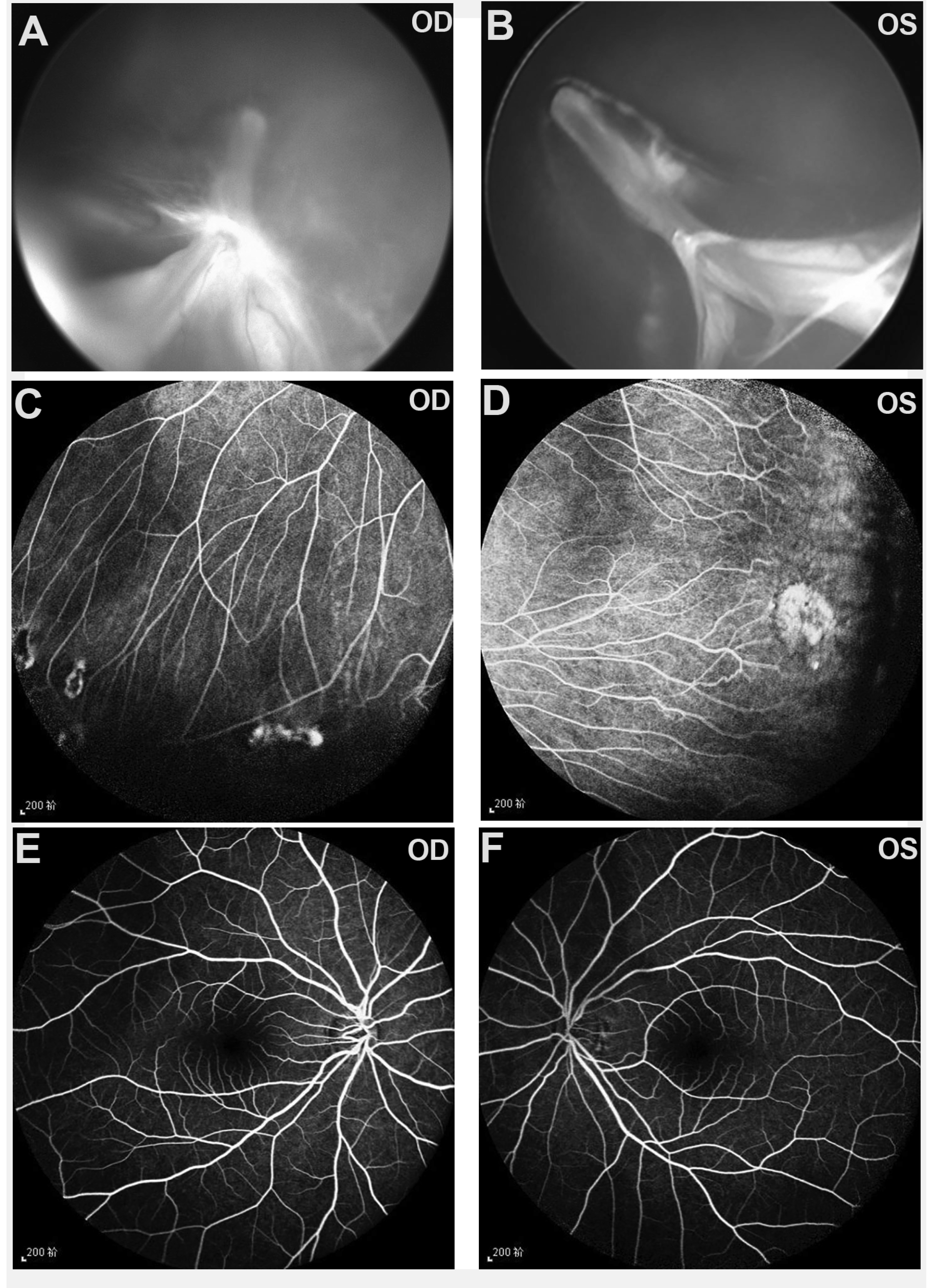
 Figure 5 of
Tian, Mol Vis 2019; 25:60-69.
Figure 5 of
Tian, Mol Vis 2019; 25:60-69.