Figure 3. Fundus and angiographic images of family B with familiar exudative vitreoretinopathy. A, B: Fundus photographs of the proband show the peripheral avascular area in the right eye and falciform retinal fold in the
left eye. Angiography was not performed for the proband because of his parents’ unwillingness. C, D: The angiographic images of his affected father show straightened vessels and an avascular area in the peripheral retina.
E, F: His mother has normal retinal vasculature demonstrated with angiography.
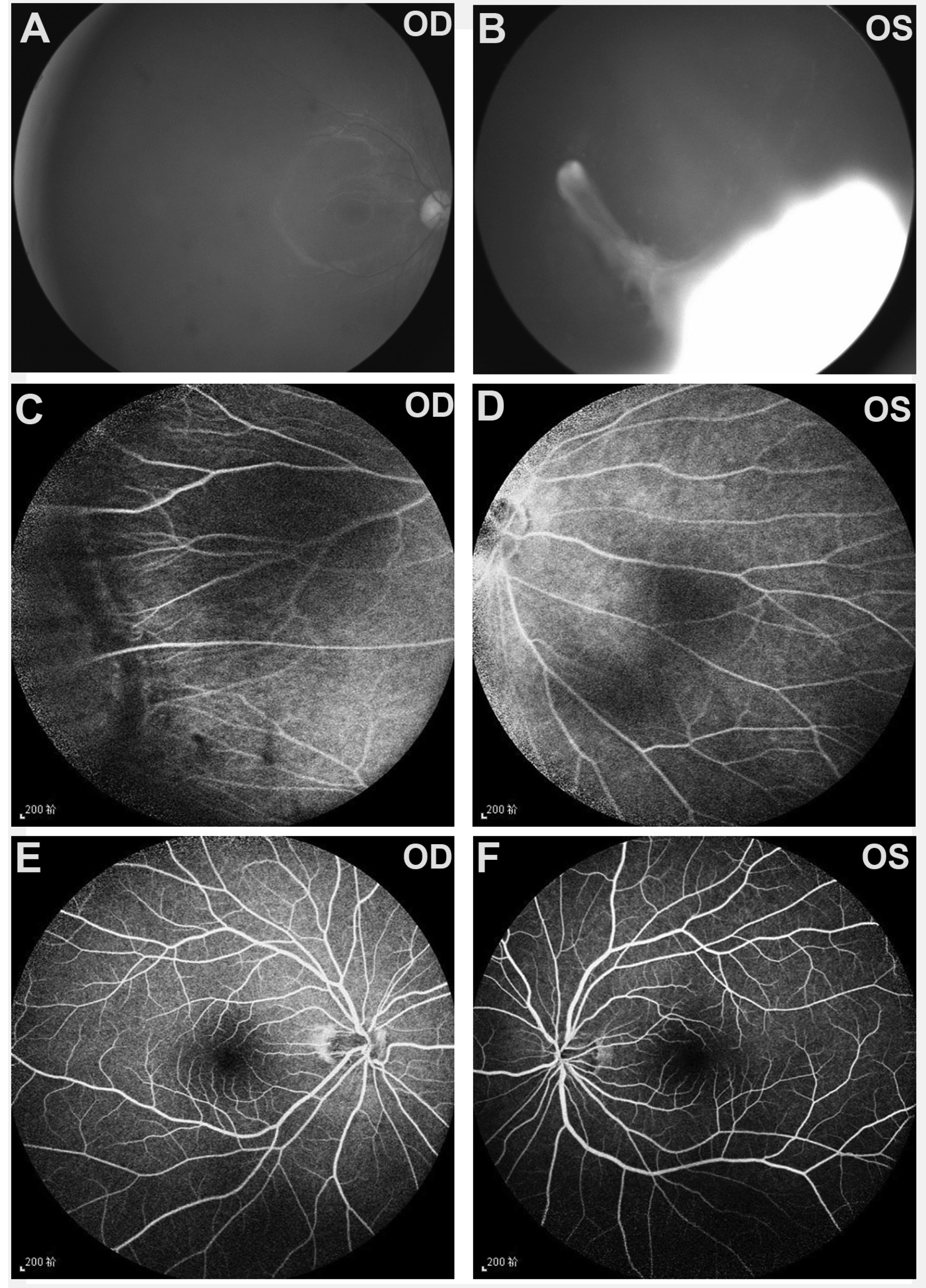
 Figure 3 of
Tian, Mol Vis 2019; 25:60-69.
Figure 3 of
Tian, Mol Vis 2019; 25:60-69.