Figure 2. Fundus and angiographic images of family B with familiar exudative vitreoretinopathy. A, B: Color photographs and an angiographic image of the proband show a disappeared anterior chamber in the right eye and aberrant
vessels with an avascular area in the peripheral retina of the left eye. A control picture of a normal anterior chamber from
another individual is provided as reference (bottom right corner in A). C–F: His mutation-carrying father is asymptomatic with a peripheral avascular area and abnormal vessels in the left eye. G, H: His mother has normal retinal vasculature demonstrated with angiography.
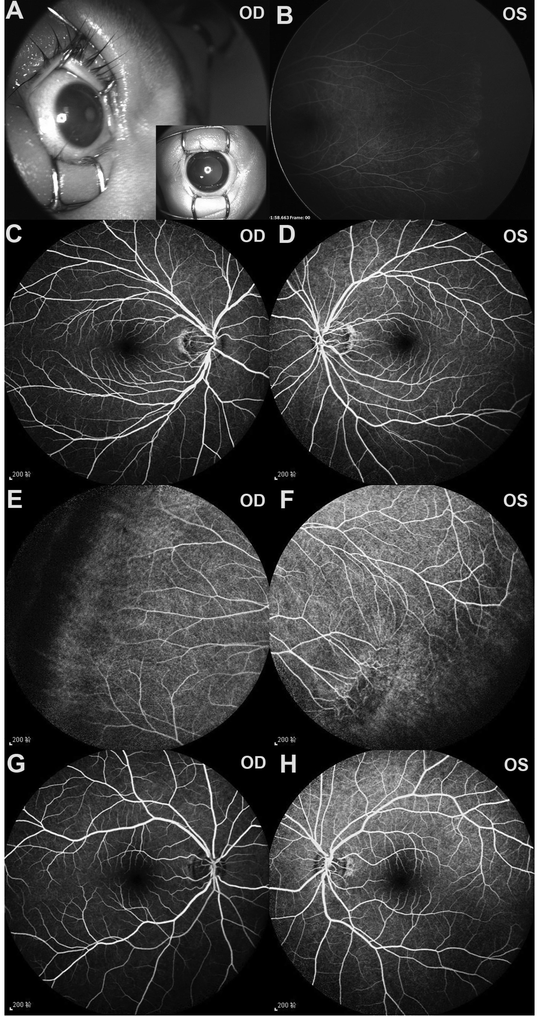
 Figure 2 of
Tian, Mol Vis 2019; 25:60-69.
Figure 2 of
Tian, Mol Vis 2019; 25:60-69.