Figure 2. Confocal microscopy cross-sectional images showing a bright band of fluorescently labeled transferrin outside the lens capsule
from a young donor (left); in contrast, fluorescently labeled transferrin is seen inside the lens capsule from an older donor
(right). O: outside the lens capsule, LC: inside the lens capsule, E: epithelial cells, L: lens. The scale bar represents
10 μm.
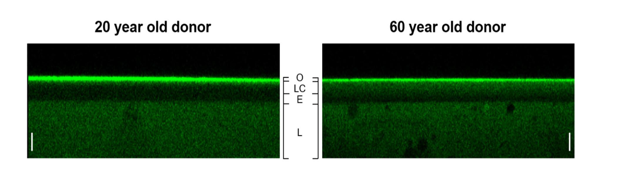
 Figure 2 of
Sueiras, Mol Vis 2019; 25:593-602.
Figure 2 of
Sueiras, Mol Vis 2019; 25:593-602.