Figure 2. IVCM images of each group on day 14 after the CXL. The control group: nerves existed in subepithelium stroma (A, arrow) while keratocytes reduced (A). Keratocytes in deeper stroma (E, I, M) and endothelial cell (Q) remained normal. 0.20% G-CXL group: visible nerves (B, arrow) and reduced keratocytes (B, F) were noticed. Needle-shaped structure existed in anterior stroma (F, arrowhead). Keratocytes in anterior (F) and mid-stroma (J) were activated. Posterior stroma (N) and endothelial cell were normal (R). 0.25% G-CXL group: nerves were visible (C, arrow). Needle-shaped structure existed in anterior stroma (G, arrowhead). Keratocytes in anterior to mid-stroma were reduced and activated (G, K). Posterior stroma (O) and endothelial cell (S) was normal. UVA-CXL group: nerves (D) and most keratocytes in anterior to mid-stroma (D, H, L) disappeared. Needle-shaped structure existed in mid-stroma (L, arrowhead). Posterior stroma (P) was normal. Endothelial cell was obvious damaged (T).
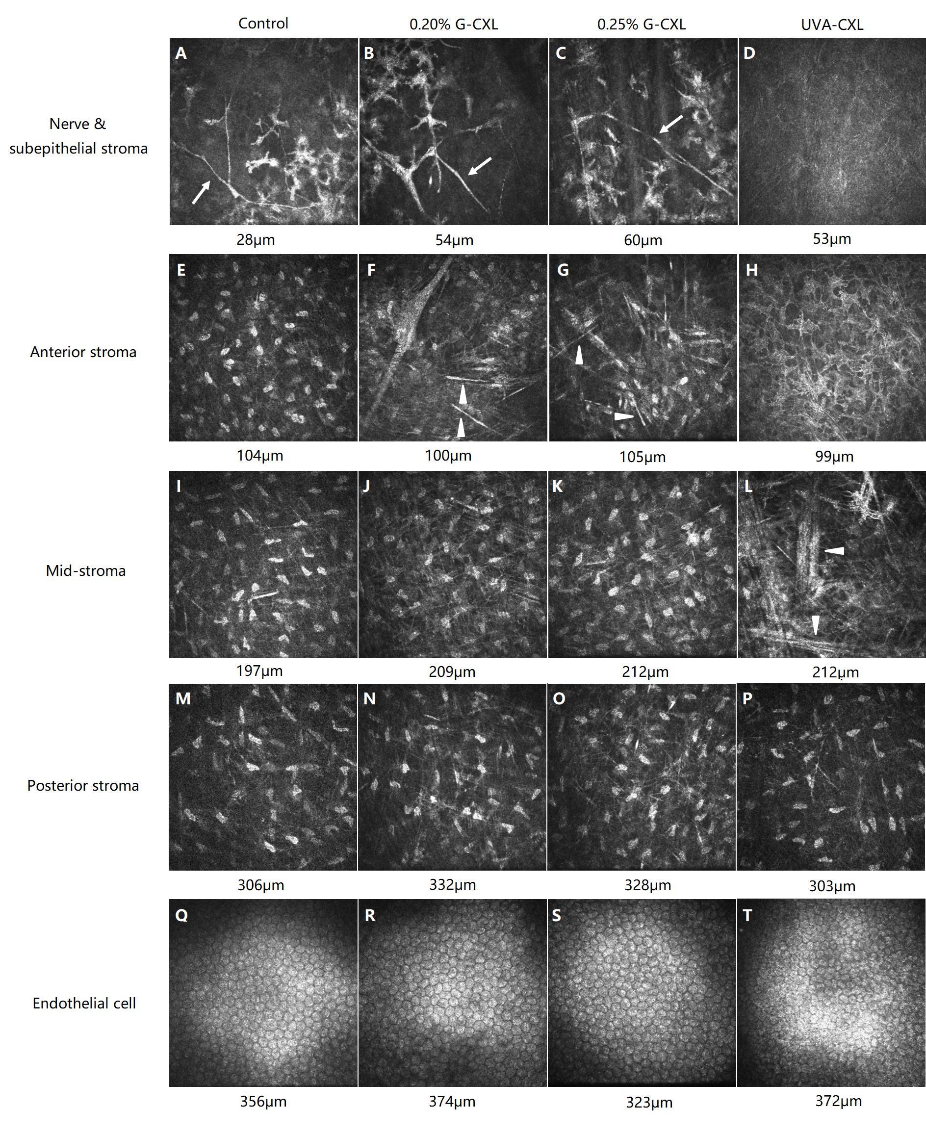
 Figure 2 of
Tang, Mol Vis 2019; 25:574-582.
Figure 2 of
Tang, Mol Vis 2019; 25:574-582.