Figure 1. ASOCT images of each group on day 14. A: DL in 0.20% G-CXL group, located in the anterior stroma(arrows). The deepest position was on the superior edge of pupil
(arrowhead). B: DL in 0.25% G-CXL group, located in anterior-mid stroma (arrows). The deepest position was on the nasal edge of pupil. C: DL in UVA-CXL group (arrows). The deepest position was in the cornea center (arrowheads). D: The control group, no DL was
noticed.
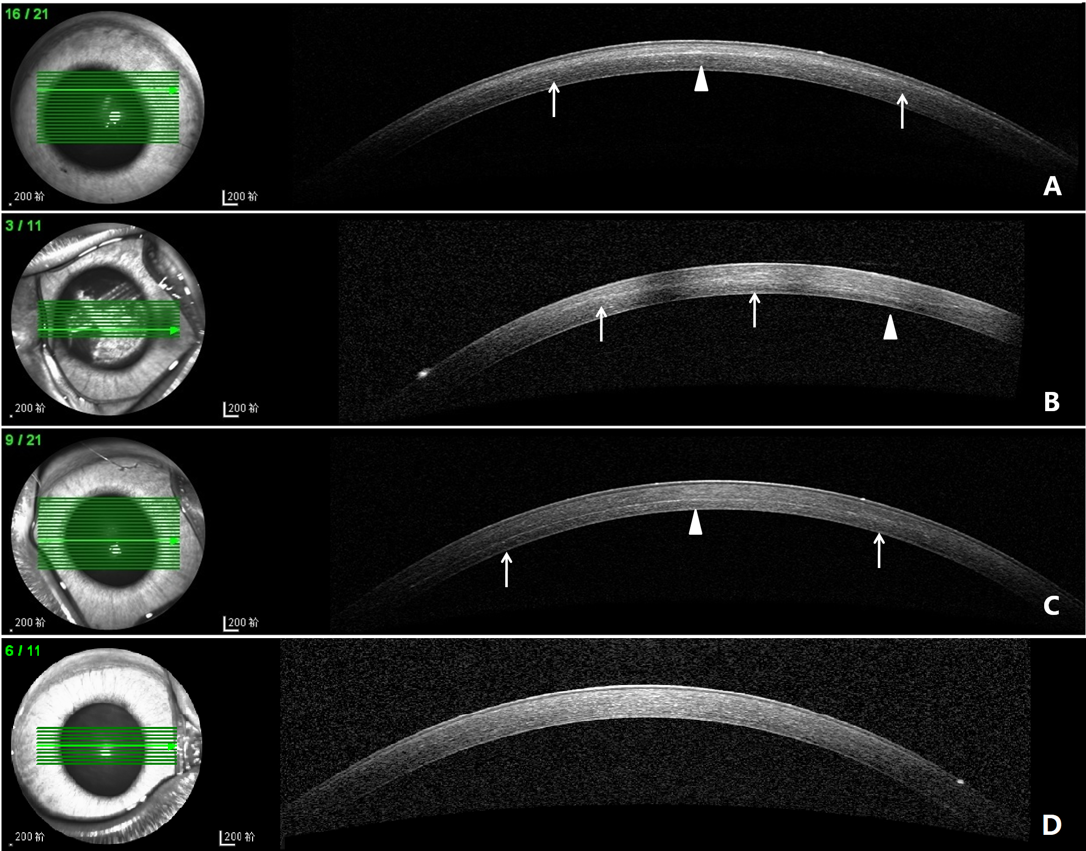
 Figure 1 of
Tang, Mol Vis 2019; 25:574-582.
Figure 1 of
Tang, Mol Vis 2019; 25:574-582.