Figure 4. Optical coherence tomographic (OCT) images along the horizontal meridian. An example of a normal 19-year-old woman is shown
at the bottom. In all cases, a thinning of the nerve fiber layer (NFL) and ganglion cell layer (GCL) was observed between
the optic disc and the fovea (asterisk).
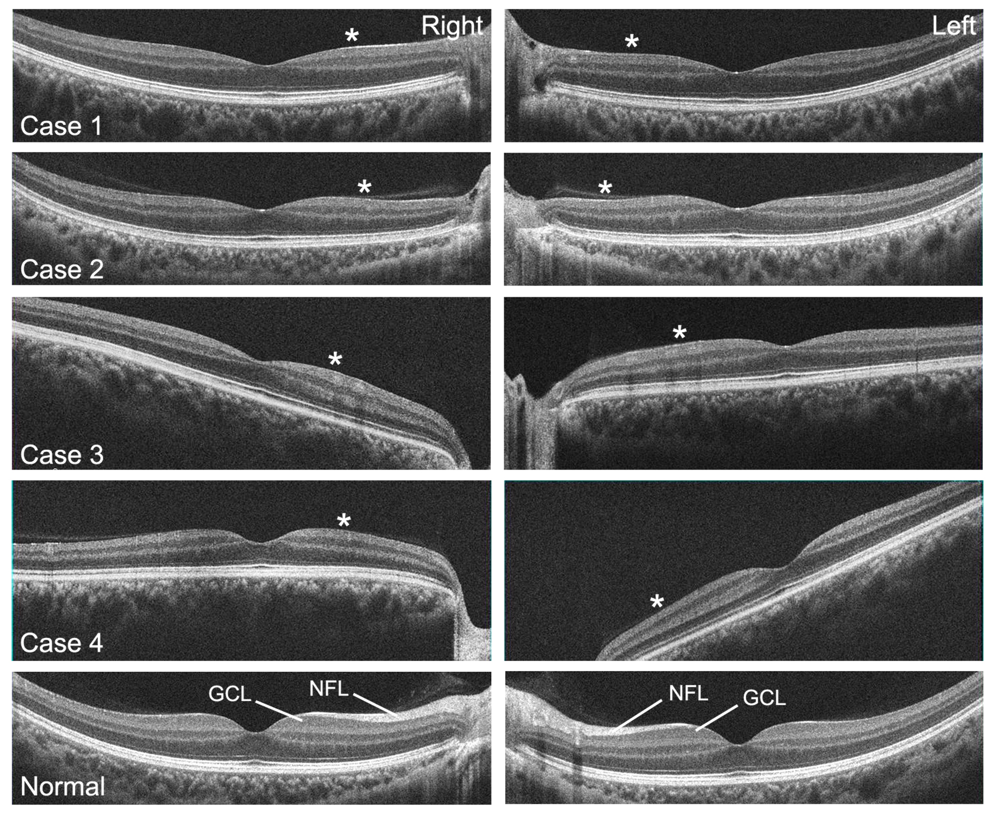
 Figure 4 of
Maeda-Katahira, Mol Vis 2019; 25:559-573.
Figure 4 of
Maeda-Katahira, Mol Vis 2019; 25:559-573.