Figure 3 of
Maeda-Katahira, Mol Vis 2019; 25:559-573.
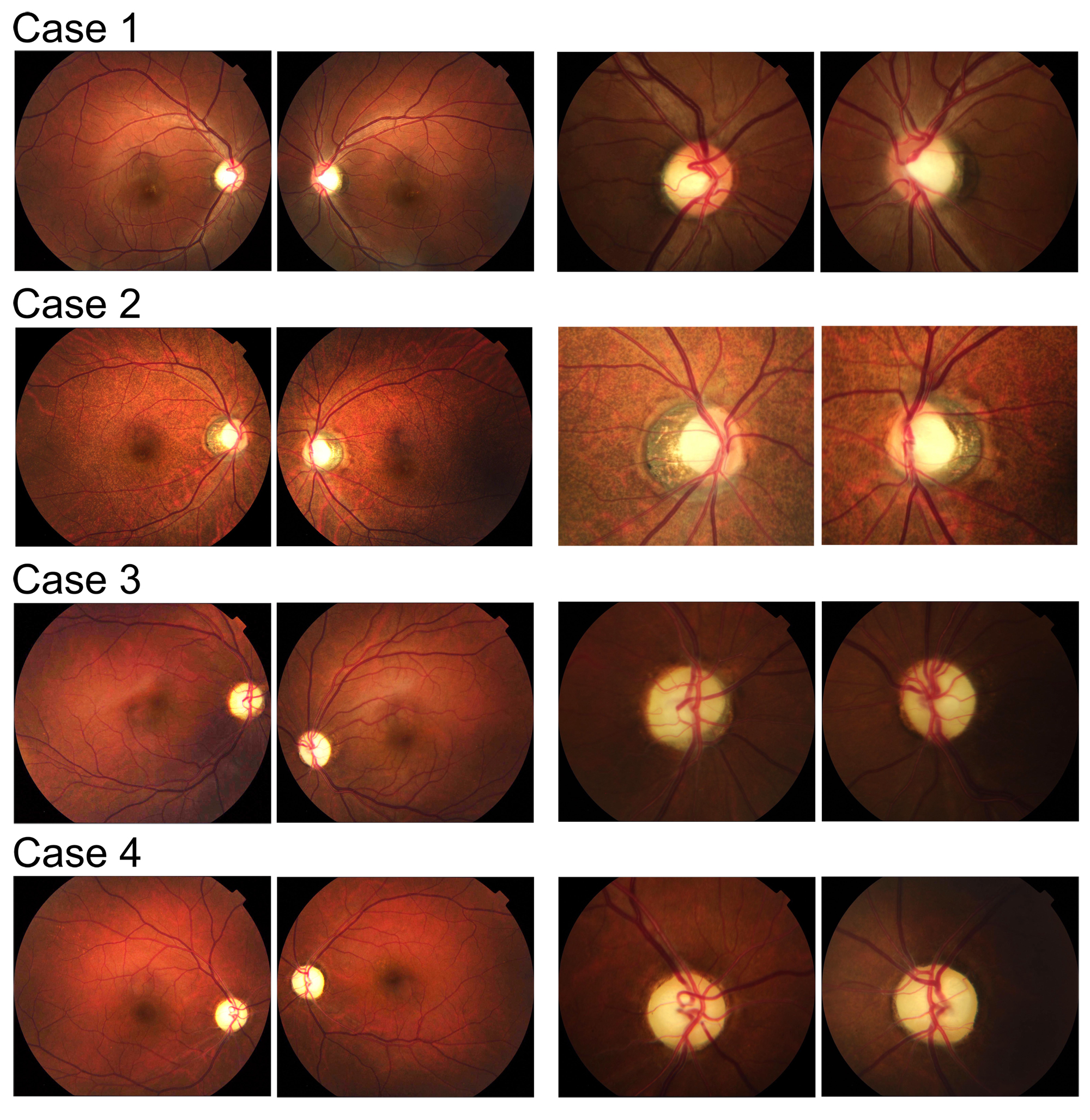
Figure 3.
Fundus photographs of the four DOA-plus patients with enlarged optic discs in the left columns. Diffuse or temporal pallor of the optic disc was present in all cases.
 Figure 3 of
Maeda-Katahira, Mol Vis 2019; 25:559-573.
Figure 3 of
Maeda-Katahira, Mol Vis 2019; 25:559-573.