Figure 1. RPE cell morphology and identity. RPE cultures without any treatment were stained with antibodies against RPE65 (A), zonula occludens 1 (ZO-1) (C), and cellular retinaldehyde-binding protein (CRALBP) (E), as described in the experimental procedure section. The corresponding bright-field images are shown in the right column
(B, D, F).
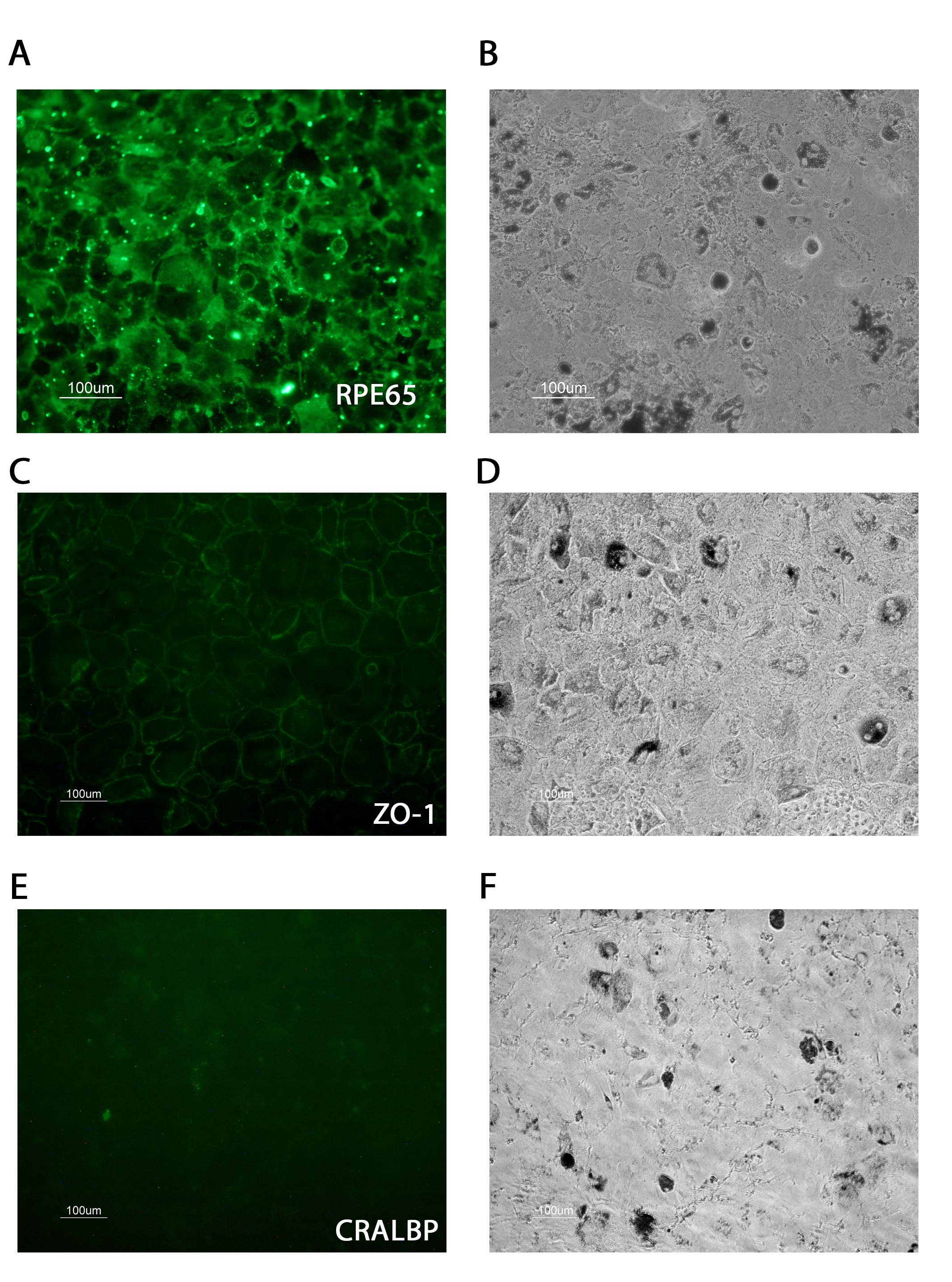
 Figure 1 of
López, Mol Vis 2019; 25:546-558.
Figure 1 of
López, Mol Vis 2019; 25:546-558.