Figure 5. Non-uniform distribution of GVs across the circumference of the canal. A: Giant vacuoles (GVs) present at the endothelial cell lining of the inner wall of Schlemm’s canal (SC). (I) A portion of
the vacuoles did not possess any intracellular pores, while some of the vacuoles had pores opening (II) toward the juxtacanalicular tissue (JCT), (III) or toward the lumen of the canal (IV), or toward the JCT and
SC. White arrows indicate the apical and basal pores. B, C: The type and distribution of GVs are non-uniform along the circumference of SC.
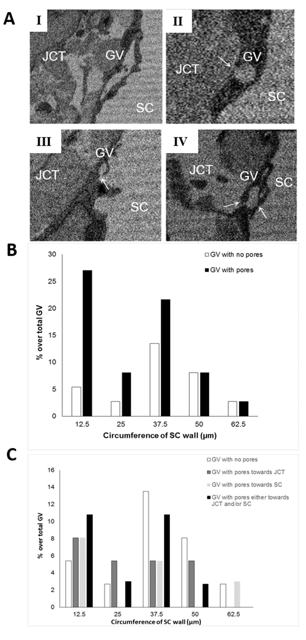
 Figure 5 of
Koudouna, Mol Vis 2019; 25:517-526.
Figure 5 of
Koudouna, Mol Vis 2019; 25:517-526.