Figure 3. Transmission electron microscopy of human TM. A: The endothelial lining of the inner wall of Schlemm’s canal (SC) is indicated by the arrowheads. A giant vacuole (GV) is
shown. B: The juxtacanalicular tissue (JCT) lies adjacent to the inner wall of SC and next to it the (C) trabecular meshwork (TM) beams are found. Scale bar=5 µm.
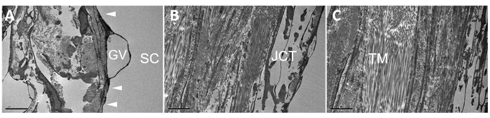
 Figure 3 of
Koudouna, Mol Vis 2019; 25:517-526.
Figure 3 of
Koudouna, Mol Vis 2019; 25:517-526.