Figure 1. Serial block face scanning electron microscopy (SBF-SEM) of the human TM. A: Light microscopy of toluidine blue-stained meridional section through corneal limbus showing Schlemm’s canal (SC), juxtacanalicular
tissue (JCT), and trabecular meshwork (TM). A giant vacuole (GV, arrow) is visible on the inner wall of SC. Scale bar, 50
μm. B: SBF-SEM was conducted in a selected area of 4,096 × 4,096 pixels with magnification equating to 27 nm/pixel, and data sets
of up to 500 images were acquired of the block face, renewed by successive slicing at 125 nm. An area of 1,588 × 1,844 pixels
was selected for this study, shown in the boxed region. C: An isolated view of the boxed region shown in B. See also Appendix 1. SC = Schlemm’s canal; JCT = Juxtacanalicular tissue; N = nucleus; ES = empty spaces.
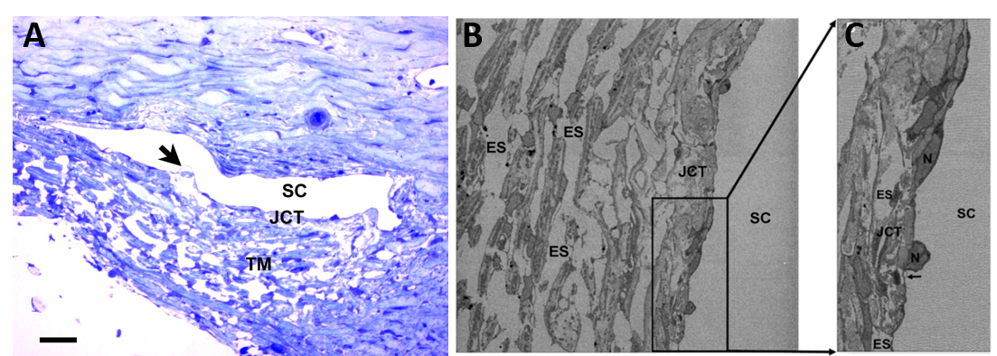
 Figure 1 of
Koudouna, Mol Vis 2019; 25:517-526.
Figure 1 of
Koudouna, Mol Vis 2019; 25:517-526.