Figure 5. Intratumor heterogeneity analysis. A: Tumors were subdivided into circular 2.0 mm diameter sections for analysis of differences in BAP-1 expression, PAS density,
and presence of VM on an intratumor region level. In this example, a region at the base of a tumor (b and c) is compared to
a region at the apex of the tumor (D and E). B: In the former, BAP-1 expression can be seen in approximately 30% of tumor cell nuclei. C: In the periodic acid-Schiff (PAS) stain from the corresponding tumor region, patterns of vasculogenic mimicry (VM) are identifiable,
including a closed loop (asterisk). D: In the region at the apex of the tumor, BAP-1 expression is higher. E: No patterns of VM can be seen. F: Bar plot and cross tabulation comparing BAP-1 expression and the presence of VM in 212 intratumor regions. Regions with
low BAP-1 expression correlated to regions with VM and vice versa (Fisher’s exact p<0.0001). G: Box plot showing PAS density in intratumor regions with low versus high BAP-1 expression (Mann–Whitney U test p<0.0001). H: Box plot showing PAS density in intratumor regions with and without VM (Mann–Whitney U test p<0.0001). I: Box plot showing the number of CD68 positive cells in regions with and without VM (Mann–Whitney U test p=0.01). Error bars represent 95% confidence interval. °=outliers, *=extreme outliers. Outlier number identifying consecutive intratumor region 1–212. Scale bars: a: 5 mm. b to e:
100 μm.
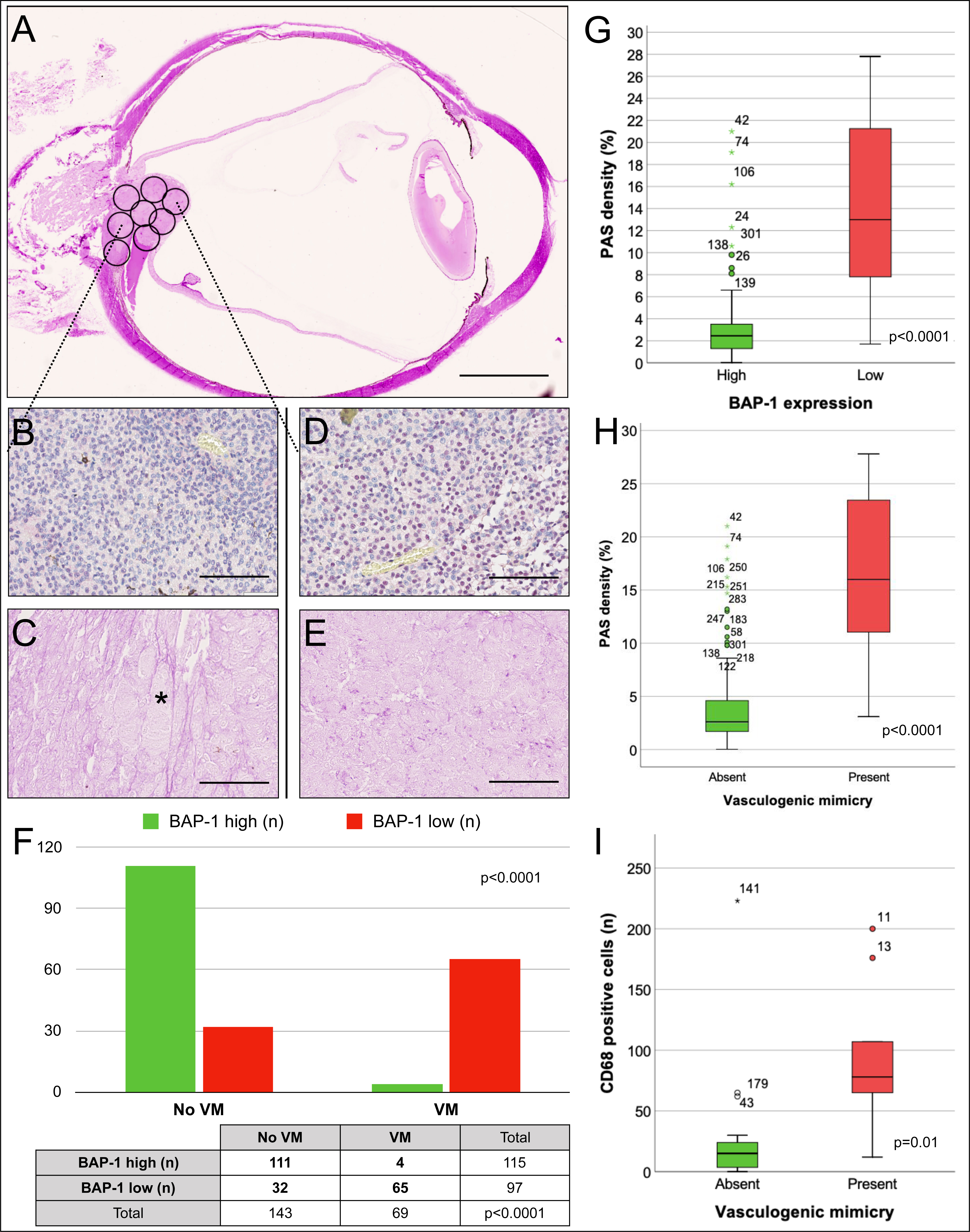
 Figure 5 of
Stålhammar, Mol Vis 2019; 25:502-516.
Figure 5 of
Stålhammar, Mol Vis 2019; 25:502-516.