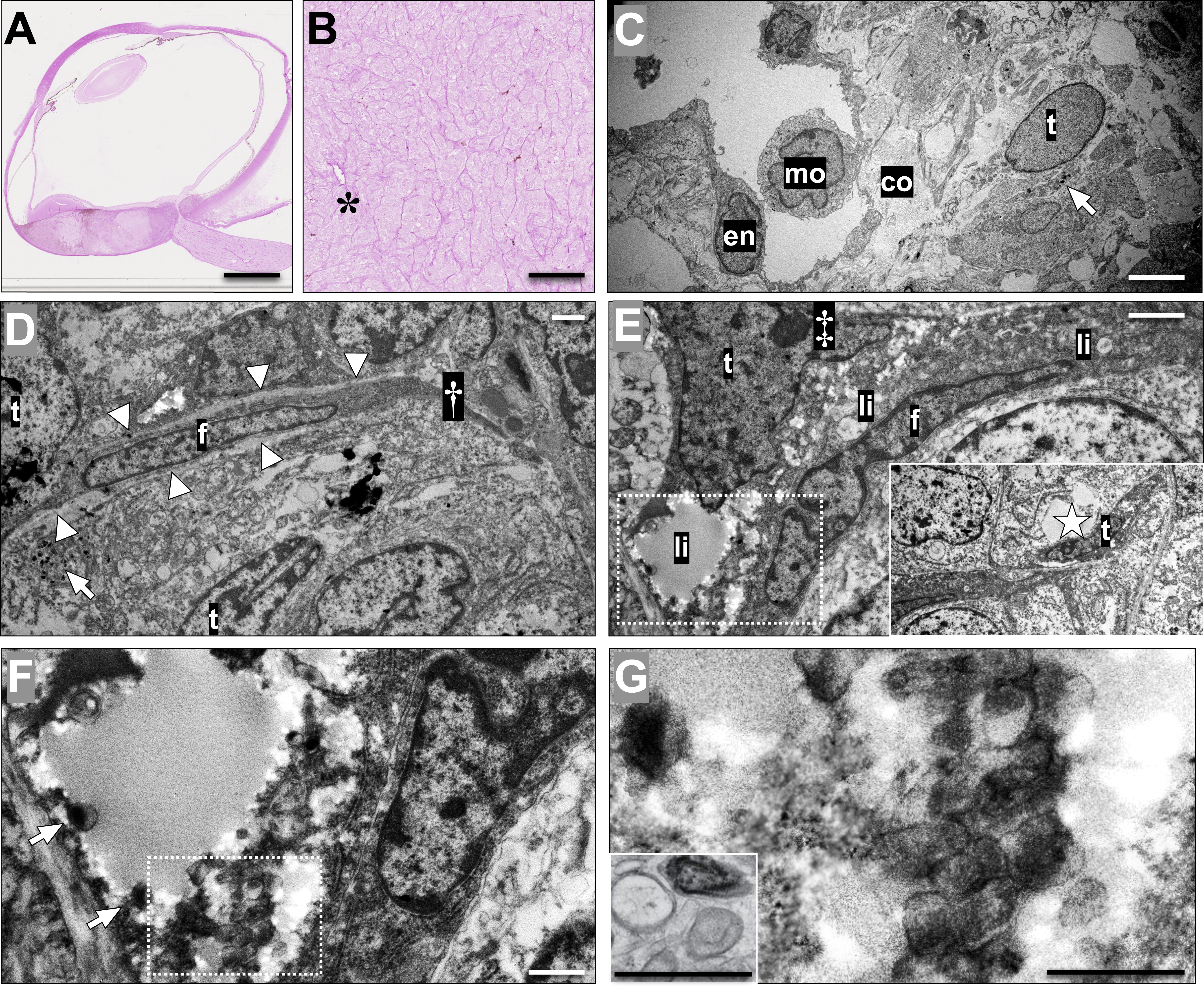
Figure 3. Choroidal vessels and vasculogenic mimicry with bright-field microscopy and transmission electron microscopy.
A: Tissue section from an enucleated eye with a posterior choroidal melanoma, stained with periodic acid-Schiff (PAS) without
hematoxylin counterstain.
B: In higher magnification, patterns of vasculogenic mimicry including closed loops and networks surrounding packets of melanoma
cells can be identified, along with a blood vessel (asterisk).
C: In transmission electron microscopy of the same tumor, a blood vessel can be identified with a lumen containing a monocyte
(mo, marking the nucleus) and a lining of endothelium (en). The vessel is surrounded by bundles of collagen (co) and a tumor
cell (t) with cytoplasmic melanosomes (arrow).
D: In another area of the tumor, a fibroblast (f) is wedged between tumor cells (t), the latter being identifiable by cytoplasmic
melanosomes (arrow). The fibroblast is associated with a fibrous septum extending outside the cytoplasm (†), and is lined
on both sides by a light, thin basal lamina (arrowheads). However, no potential lumen or fluid can be identified in this area.
E: In contrast, in yet another tumor area, a tumor cell (t), characterized by a large nucleus with a nucleolus (‡), can be
seen sharing a potential space full of debris and lipid droplets (li) with a fibroblast (f). No basal lamina can be identified
between the fibroblast and the tumor cells. The potential space extends to a larger sinus (insert, sinus marked by a star),
which is lined by a flattened tumor cell (t), recognizable by its large nucleolus and coarse chromatin.
F: Magnification of the area within the dashed box in
E. Note that melanosomes can be seen in the cytoplasm of the tumor cell (arrows).
G: Magnification of the area within the box in
F. Note that in addition to the mature melanosomes, ovoid premalonosomes can be identified (arrowheads), comparable to an example
of premature and mature melanosomes in a line of MNT-1 skin melanoma cells, fixed by high-pressure freezing (insert, modified
from Raposo et al. [
27]; reprinted with permission from Springer Nature). This tumor was classified as 1a by gene expression profiling, and had
retained nuclear BAP-1 expression. Still, the patient developed metastases 10 months after enucleation. Scale bars: a: 5 mm.
b: 200 μm. c: 5 μm. d, e: 2 μm. f, g: 1 μm.
 Figure 3 of
Stålhammar, Mol Vis 2019; 25:502-516.
Figure 3 of
Stålhammar, Mol Vis 2019; 25:502-516.