Figure 4. Assessment of the effect of antioxidant treatment on photic retinal injury in mice. Mice treated with the G2, G3, and G4 formulas
(see the Methods section for details) were evaluated for oxidative injury (A), apoptosis (B), outer nuclear layer thickness (C, D), and electroretinography (ERG) response (E, F). Oxidative injury was evaluated with measurement of 4-hydroxynonenal (4-HNE) antibody label tissue. A reduced level of HNE
antibody label tissue was detected 7 days after the exposure to light, compared to the vehicle-treated group (n=8, one-way
ANOVA Dunnett, *p≤0.05, A). Apoptosis was assessed with measurement of the number of activated caspase 3+ cells, which was similar across the treatment and vehicle groups (one-way ANOVA, p>0.05, B). The level of oxidative stress and the number of caspase 3+ cells were normalized to the control group (not exposed to light). None of the treatments conferred a protective effect on
the photoreceptors, as shown with the measurements of the number of nuclei in the outer nuclear layer (one-way ANOVA, p>0.05,
C and D), and with ERG recording (one-way ANOVA, p>0.05, E and F). The measurement of ONL thickness was normalized to the vehicle group.
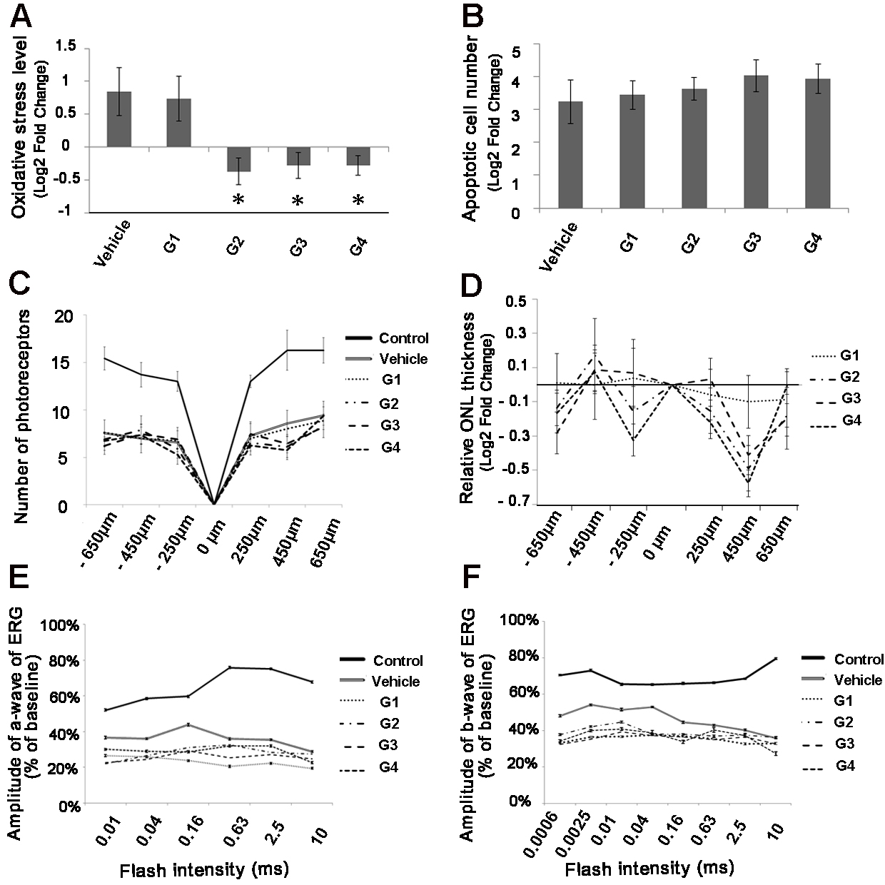
 Figure 4 of
Elbaz-Hayoun, Mol Vis 2019; 25:479-488.
Figure 4 of
Elbaz-Hayoun, Mol Vis 2019; 25:479-488.