Figure 3. Establishing the model of photic retinal injury. Exposure of BALB/c mice to bright light of 8000 lux intensity for 3 h led
to 50% loss of photoreceptors 7 days after light injury at different distances from the optic disc (−2-fold, unpaired t test,
*p=0.01, **p=0.003, ***p=0.0003, A: Histological sections, stained with 4′,6- diamidino-2-phenylindole (DAPI; blue signal), showed reduction of the ONL thickness
after photic injury (B and C) compared to that of the control group (D and E). An increase in the oxidative stress level was observed 7 days after the light injury (1.63-fold, unpaired t test, *p=0.009,
F and G), compared to the control mice not exposed to light (H), as shown with the 4-hydroxynonenal (4-HNE) antibody label tissues (green signal). The fluorescence intensity of the 4-HNE
antibody label tissue was measured using ImageJ software, and the negative control of 4-HNE antibody label tissue value was
subtracted. Recruitment of cd11b+ cells (red signal, arrows) was observed 7 days after light injury (I), compared to the control group (J). GCL, ganglion cell layer; INL, inner nuclear layer; ONL, outer nuclear layer.
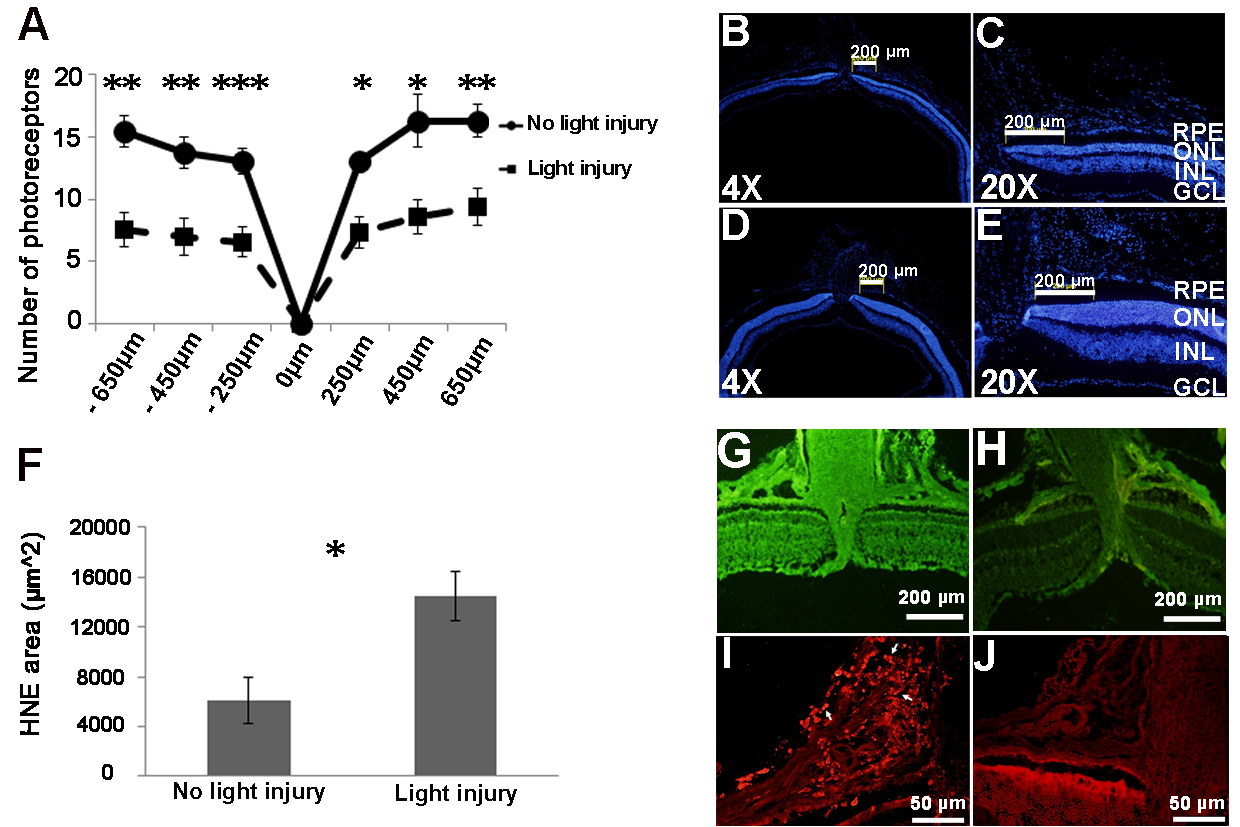
 Figure 3 of
Elbaz-Hayoun, Mol Vis 2019; 25:479-488.
Figure 3 of
Elbaz-Hayoun, Mol Vis 2019; 25:479-488.