Figure 1. In vitro assessment of the effect of antioxidant supplements on the angiogenic properties of M1 and M2a hMDMs. Four antioxidant
treatments (G1–G4; see the details in the Methods section) were added to the culture medium of M1 (n=7) and M2a (n=8) human
monocyte-derived macrophages (hMDMs). The addition of untreated culture medium served as control (G5). Choroid tissue was
cultured for 8 days with the supernatants from M1 (A) and M2a (B) hMDMs treated with the different supplements. The sprouting area was calculated using ImageJ software (repeated-measure
mixed-effect model, *p≤0.05). C: Representative image from control is shown. D: The G3-treated medium culture of M2a macrophages is shown.
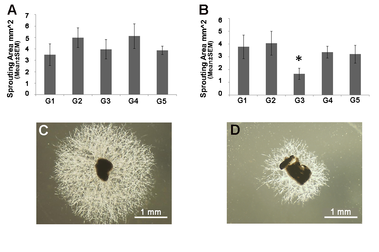
 Figure 1 of
Elbaz-Hayoun, Mol Vis 2019; 25:479-488.
Figure 1 of
Elbaz-Hayoun, Mol Vis 2019; 25:479-488.