Figure 4. Exercise partially preserves outer nuclear layer in the I307N Rho retinal degeneration. I307N Rho degeneration was induced in active and inactive mice. A week later, the mice were euthanized, and ocular sections stained
with hematoxylin and eosin (H&E). A: Representative H&E images of retina sections from each treatment group are from a region of 250–750 μm from the optic nerve.
Examples of complete sections are shown in Appendix 5. B: Nuclei were counted in six discrete regions of the retinal sections starting at 250 µm from the optic nerve head and extending
every 500 µm outward along the dorsal or superior (positive values on abscissa) and ventral periphery or inferior (negative
numbers on abscissa); spacing and counting overlay templates are shown in Appendix 5. The inactive, induced mice (red) showed
statistically significant loss of nuclei at four distances from the optic nerve head compared to the uninduced group (black);
**p<0.01 and ****p<0.0001 versus the inactive, uninduced group. However, the induced mice that were active (green) exhibited
mean nuclei counts statistically indistinguishable from those of the inactive, uninduced group (black) throughout the length
of the retina. In the region of 250–750 μm superior and inferior from optic nerve, the nuclei counts were statistically greater
in the active versus inactive induced mice; **p<0.01 and *p<0.05, comparisons as shown in the figure. All comparisons tested
with two-way ANOVA with the Newman-Keuls multiple comparisons test. n=10–14 retinal images/group (five to seven mice/group).
Error bars represent standard error of the mean (SEM).
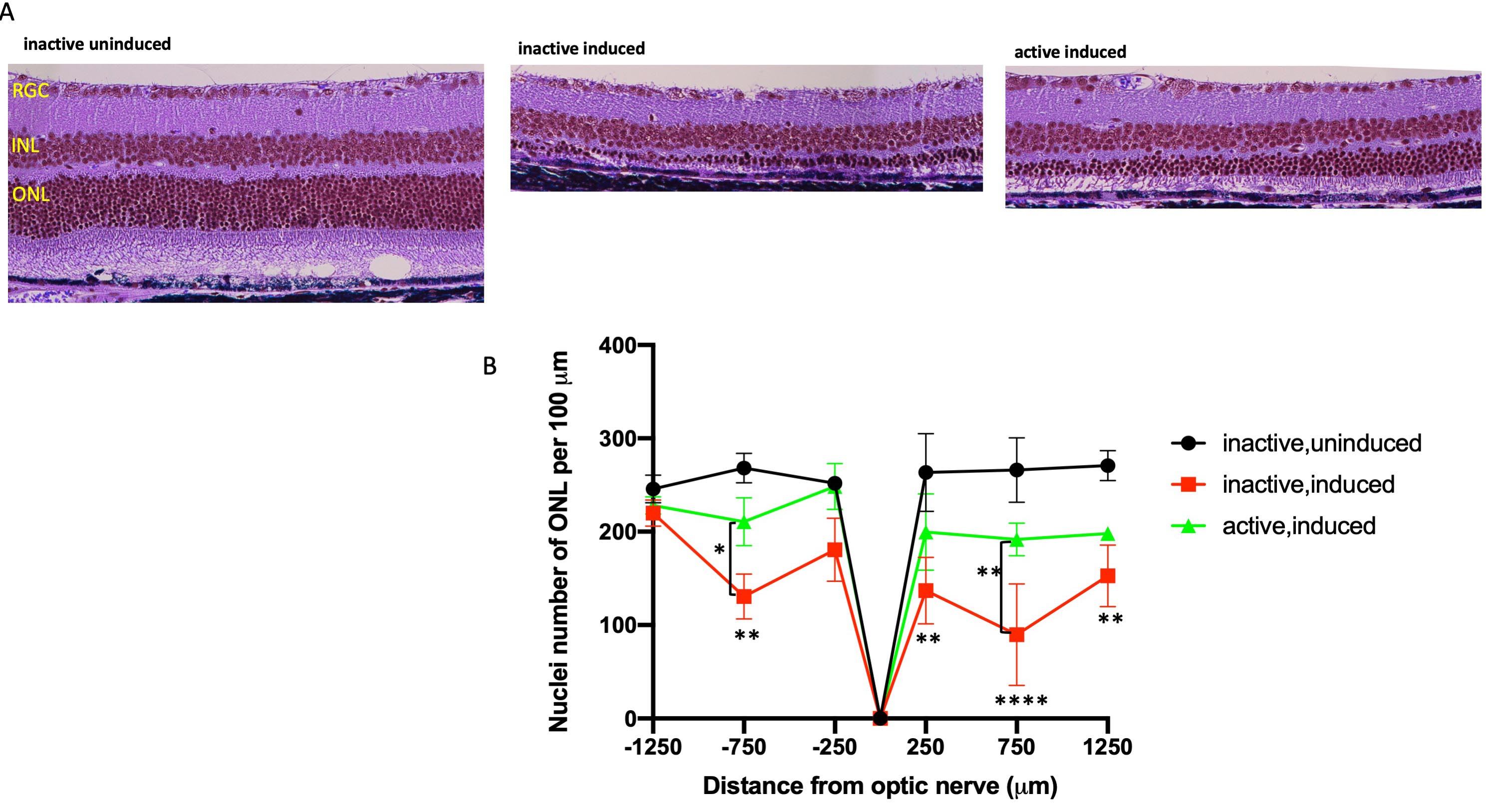
 Figure 4 of
Zhang, Mol Vis 2019; 25:462-476.
Figure 4 of
Zhang, Mol Vis 2019; 25:462-476.