Figure 3. Exercise partially preserves photoreceptor layer thickness and total retinal thickness following induction of I307N Rho degeneration. Photoreceptor thickness (A, C) and retinal thickness (B, D) from I307N Rho mice at 1 (A, B) and 4 weeks (C, D) after degeneration was induced. The mice with inactive running wheels and exposed to 6,000 lux light for 5 min (red bar)
exhibited losses in the thickness of the photoreceptor and retina layers, whereas the induced mice with active running wheels
(green bar) exhibited statistically significant preservation of layer thickness. E. Representative fundus images and corresponding optical coherence tomography (OCT) images from each group. ap<0.05 versus all other groups. Other comparisons as indicated in the figure; **p<0.01, ***p<0.001 with one-way ANOVA with
the Newman-Keuls multiple comparisons test. n=5–7 mice/group. Error bars represent standard error of the mean (SEM).
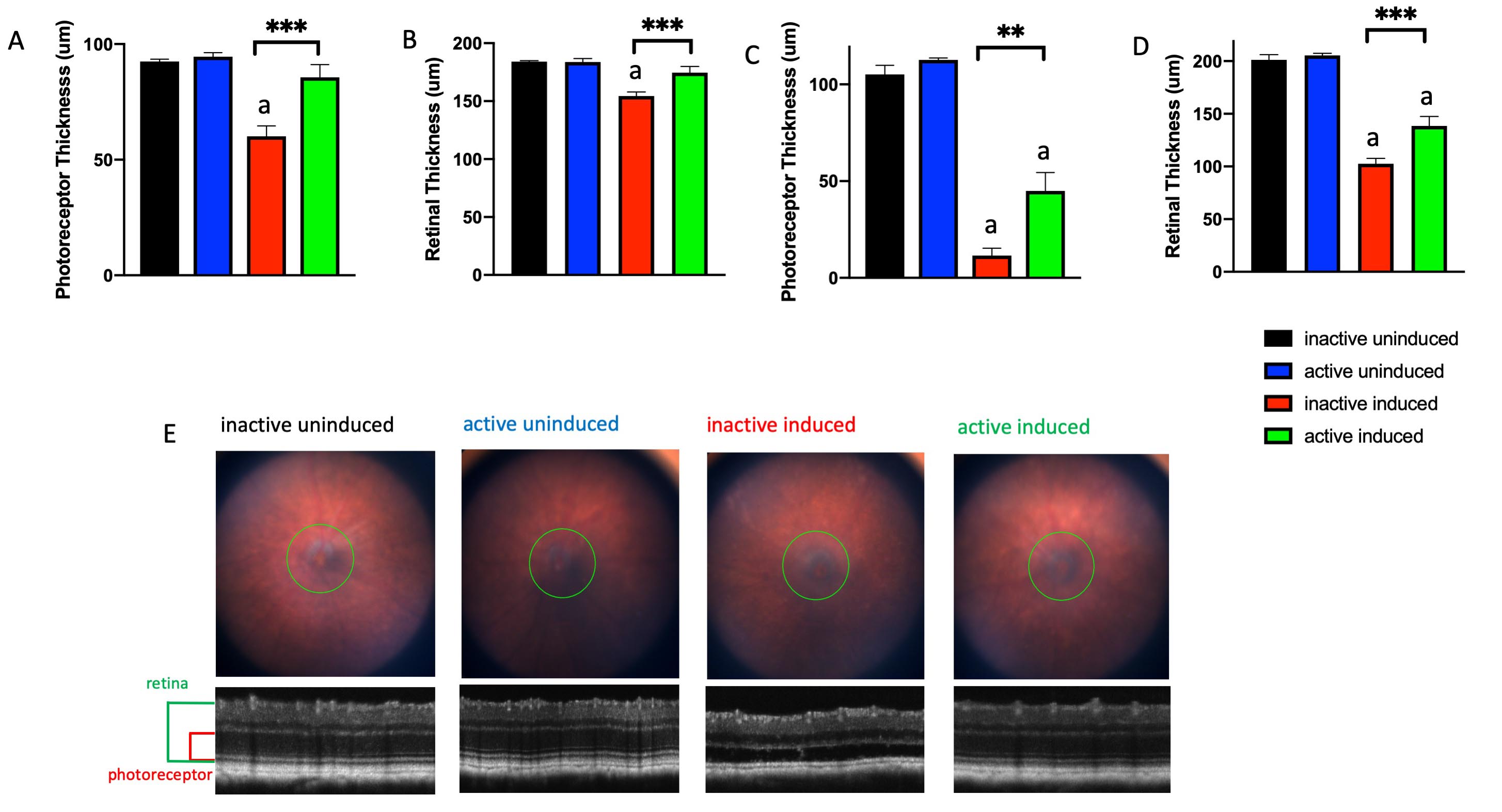
 Figure 3 of
Zhang, Mol Vis 2019; 25:462-476.
Figure 3 of
Zhang, Mol Vis 2019; 25:462-476.