Figure 1. Exercise partially preserves retinal function in the I307N Rho mouse. Scotopic electroretinogram (ERG) a-wave (A, C) and b-wave (B, D) mean amplitudes from I307N Rho mice at 1 (A, B) and 4 weeks (C, D) after degeneration was induced. Mice with inactive running wheels and exposed to 6,000 lux light for 5 min to induce degeneration
exhibited a- and b-wave mean amplitudes (red) that were statistically significantly diminished compared to those of the uninduced
groups (blue and black). However, with the exception of the data in C, the mean ERG amplitudes of the mice undergoing retinal degeneration but with active running wheels (green) were either statistically
indistinguishable from those of uninduced mice or were statistically significantly greater than those of the inactive induced
mice. *p<0.05, **p<0.01, ***p<0.001, ****p<0.0001 with two-way ANOVA with the Newman-Keuls multiple comparisons test; comparisons
as noted in the image. See Appendix 1, Appendix 2, Appendix 3, Appendix 4 for detailed tabular results and multiple comparisons.
For each group in panels A–D, n=10–14 eyes. Error bars represent standard error of the mean (SEM).
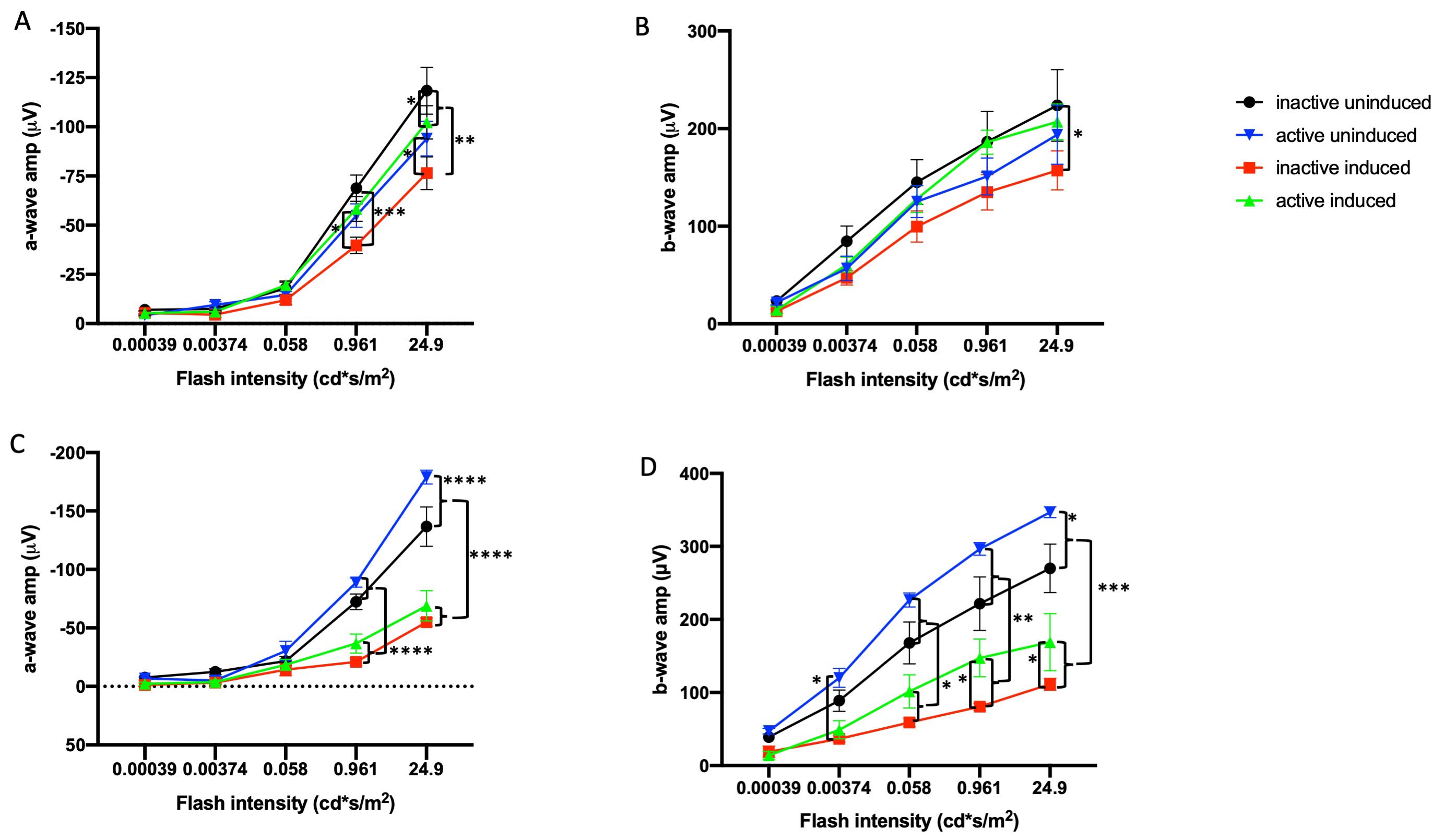
 Figure 1 of
Zhang, Mol Vis 2019; 25:462-476.
Figure 1 of
Zhang, Mol Vis 2019; 25:462-476.