Figure 6. Reorganization of collagen fibril architecture in hUMSC-treated eyes. A–D: Forward-scattered second harmonic generated (SHG) images (cyan) show that human umbilical cord mesenchymal stem/stromal
cells (hUMSC)-treated corneas (A, C) have a fibril structure more analogous to that of an uninjured eye than do the control corneas (B, D). Backscattered SHG images (magenta) show less transparent and disorganized lamellae control corneas (B, D) compared to the UMSC-treated corneas (A, C).
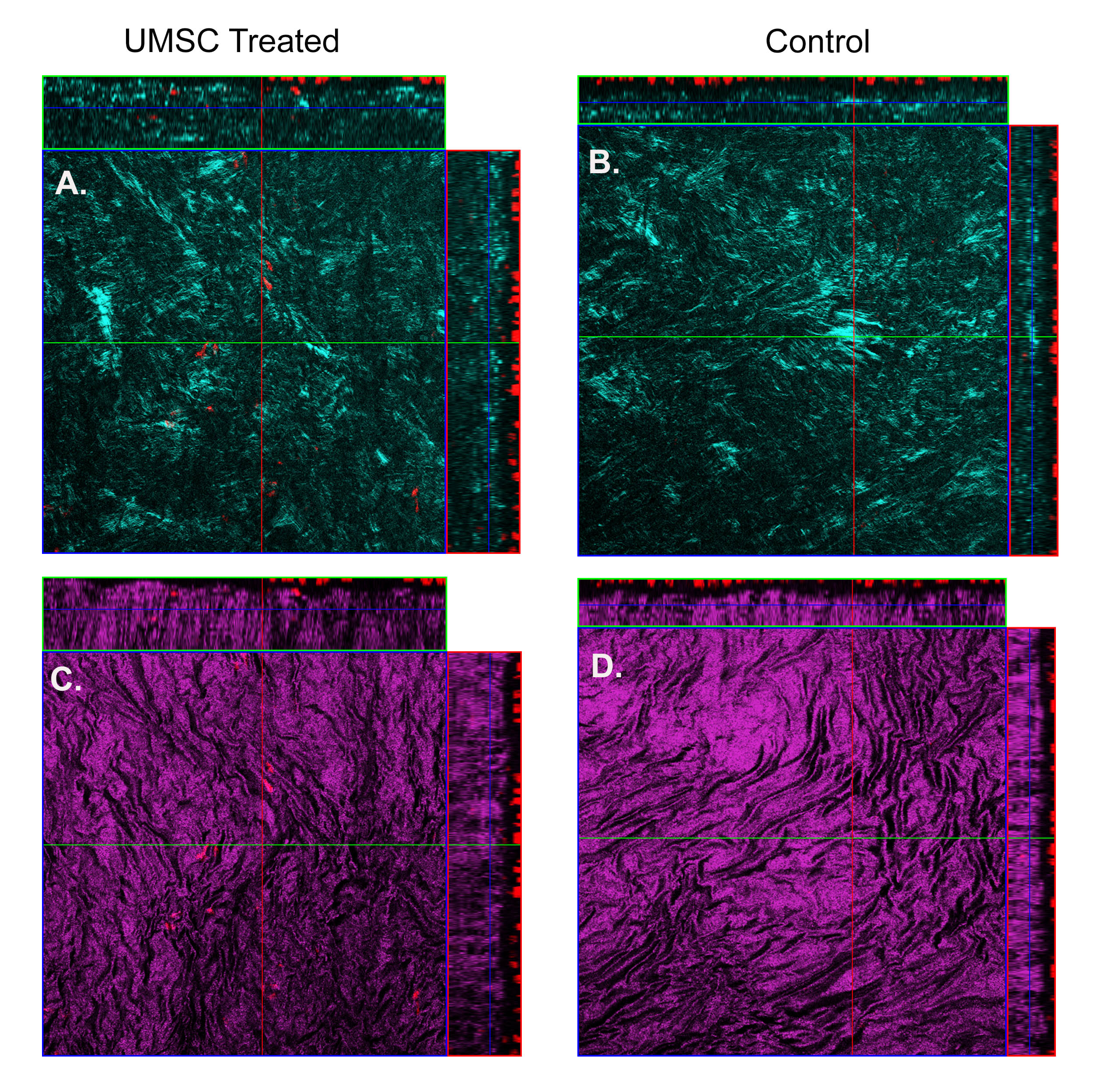
 Figure 6 of
Call, Mol Vis 2019; 25:415-426.
Figure 6 of
Call, Mol Vis 2019; 25:415-426.