Figure 2 of
Tu, Mol Vis 2019; 25:391-399.
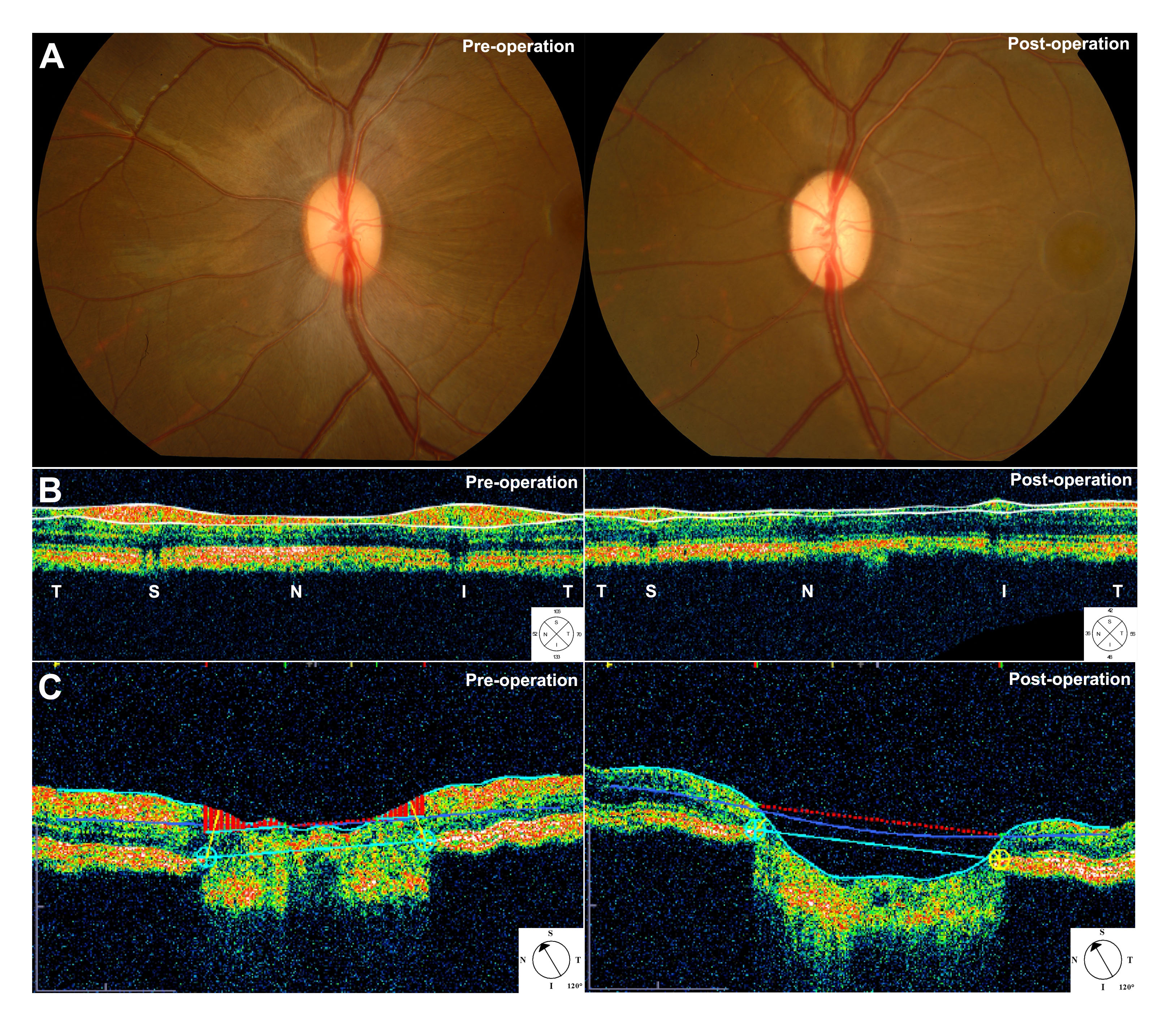
Figure 2.
A monkey model of chronic ocular hypertension was established.
A
: Fundus: The enlarged optic cup.
B
: Optical coherence tomography (OCT): The damaged retinal nerve fiber layer.
C
: OCT: The enlarged optic cup.
 Figure 2 of
Tu, Mol Vis 2019; 25:391-399.
Figure 2 of
Tu, Mol Vis 2019; 25:391-399.