Figure 3. In vivo labeling of retinal microglia with Fluorescent 4Di-10ASP. A: The whole-mount of the retinas shows the staining of the microglia (small ramified cells, white arrows) and ganglion cells
(large cell bodies, yellow arrows) after optic nerve transection and 4Di-10ASP was packed for 14 days. Scale bars: 100 μm.
Inset: higher magnification. B: Representative images from the whole-mount of the retinas show microglia staining after labeled with 4Di-10ASP for 28 days.
Scale bars: 100 μm. Inset: higher magnification. C: The retinas, which were labeled with 4Di-10ASP for 28 days, were stained with the microglial marker CD11b (red), and the
representative images were obtained with confocal microscopy. Scale bars: 50 μm.
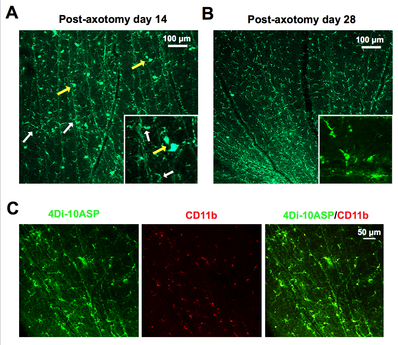
 Figure 3 of
Yuan, Mol Vis 2019; 25:359-372.
Figure 3 of
Yuan, Mol Vis 2019; 25:359-372.