Figure 2. Minocycline attenuates LPS-induced inflammation in all ocular tissues. Histopathologic sections from the normal control eyes,
and lipopolysaccharide (LPS) stimulation with or without minocycline treatment eyes were stained with hematoxylin-eosin. Representative
images from light microscopy display that the anterior chamber, iris, and ciliary body are filled with fibrin (**) and infiltrated
inflammatory cells (black arrows), and several inflammatory cells (black arrows) infiltrate into the retina in the PBS-treated
retinas. However, minocycline treatment attenuates the inflammation response in all ocular tissues (n=6 per group). Scale
bars: 100 μm for anterior chamber sections; 50 μm for iris, ciliary bodies, and retina sections.
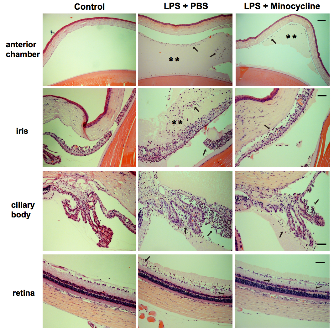
 Figure 2 of
Yuan, Mol Vis 2019; 25:359-372.
Figure 2 of
Yuan, Mol Vis 2019; 25:359-372.