Figure 4. PCR–Sanger sequencing validating the candidate EYS variants in the retinitis pigmentosa family 1 (RP-F1) and RP-F2 DNA sequencing profiles of the identified mutations (upper)
and their wild-type form (lower). Red arrows indicate the position of the mutated nucleotide. A,B: From RP-F1; C,D: from RP-F2.
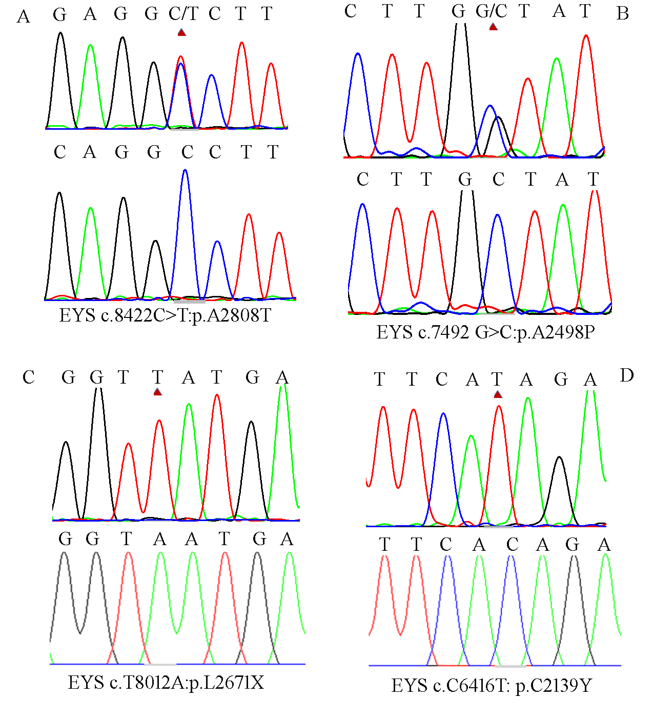
 Figure 4 of
Xiao, Mol Vis 2019; 25:35-46.
Figure 4 of
Xiao, Mol Vis 2019; 25:35-46.