Figure 2. Clinical information for the proband of retinitis pigmentosa family 1 (RP-F1). A,B: Fundus picture of the right (A) and left (B) eyes. C,D: Optical coherence tomography (OCT) scans of the right (C) and left (D) eyes. E,F: Visual fields of the right (E) and left (F) eyes. G,H: Electroretinogram (ERG) results of the right (E) and left (F) eyes.
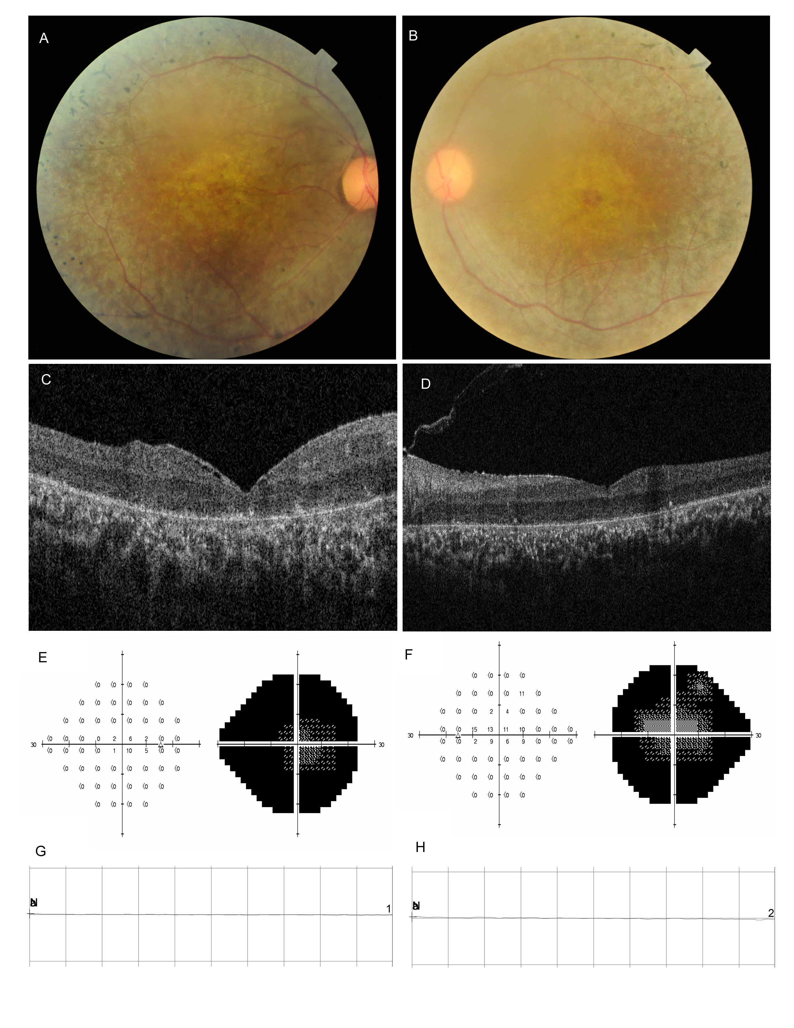
 Figure 2 of
Xiao, Mol Vis 2019; 25:35-46.
Figure 2 of
Xiao, Mol Vis 2019; 25:35-46.